
CASE REPORT
Case report of Commotio Retinae A 29-year-old woman presented with reduced vision and flickering lights in her left eye when looking up, two days following a high-velocity impact of a champagne cork ejected approximately three meters away.

Her visual acuity was 6/12+2 in the affected eye with a −1.75 D myopic shift relative to the previous refractive status (+0.50/−0.50 × 8, 6/6+) and no improvement with pinhole testing. No signs of traumatic mydriasis were observed, with both pupils equal in size, regular, round and reactive to light.
Confrontation fields and excursions were all unremarkable. Anterior segment biomicroscopy revealed mild superotemporal conjunctival ecchymosis and moderate hyperemia, along with both grade 0.5 cells and flare in the anterior chamber.
Mild lid edema and eyelid ecchymosis were also observed. There was no evidence of other anterior segment injuries, such as corneal trauma, iris tears, hyphaema, or lens opacification.
Gonioscopy showed no signs of angle recession and Goldmann applanation tonometry was 15 mmHg in each eye.
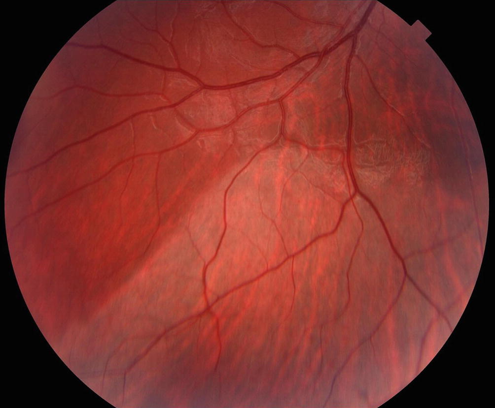
Dilated fundus examination revealed superotemporal peripheral commotio.
DISEASE of Commotio Retinae
Refer to traumatic retinopathy secondary to direct or indirect trauma to the globe. Retinopathy may be present at areas of scleral impact (coup) and or distant sites (contrecoup) including the macula.
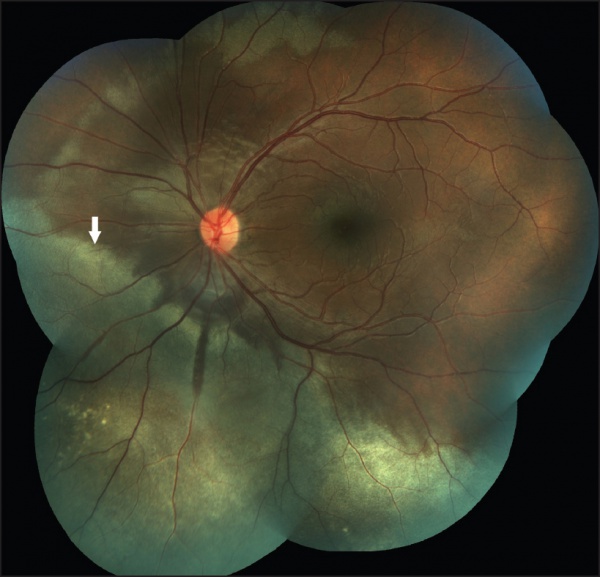
When found in the posterior pole is also referred to as Berlin’s edema.
How to get Choroida fundus explorer.
MANAGEMENT of Commotio Retinae
Patients suffering substantial ocular trauma with commotio should be followed with serial eye exams to evaluate for any additional treatable conditions.
As commotio of macula resolves, partial or full-thickness macular holes can develop.
would you have the interest to take retina images by smartphone?
Fundus photography is superior to fundus analysis as it enables intraocular pathologies to be photo captured and encrypted information to be shared with colleagues and patients.
Recent technologies allow smartphone-based attachments and integrated lens adaptors to transform the smartphone into a portable fundus camera and Retinal imaging by smartphone.

REFERENCES
- Berlin R. Sogenannten commotio retinae. So-called commotio retinae. Klin Monatsbl Augenheilkd 1873;1:42–78.
- BLANCH, R.J., UNDERSTANDING AND PREVENTING VISUAL LOSS IN COMMOTIO RETINAE, in College of Medical and Dental Sciences. 2014, University of Birmingham: Birmingham, UK. p. 581.
- Hart, J.C., and R. Blight, Commotio retinae. Arch Ophthalmol, 1979. 97(9): p. 1738.
- Ahn, S.J., et al., Optical coherence tomography morphologic grading of macular commotio retinae and its association with anatomic and visual outcomes. Am J Ophthalmol, 2013. 156(5): p. 994-1001.e1.
- Mansour, A.M., W.R. Green, and C. Hogge, Histopathology of commotio retinae. Retina, 1992. 12(1): p. 24-8.
Read more: Valsalva Retinopathy, Diabetic retinopathy,

RETINAL IMAGING BY YOUR SMARTPHONE