
Infantile capillary hemangioma is the most common orbital and periocular tumor of childhood, following a distinctive natural history — absent or barely visible at birth, growing rapidly over the following months, and then slowly involuting over subsequent years — that shapes every aspect of how these lesions are managed.
When periocular, its main clinical significance has little to do with the tumor itself and everything to do with its capacity to interfere with normal visual development if it happens to occlude the visual axis or distort the cornea during the critical early months of a child’s vision developing.
The Characteristic Growth Pattern
A capillary hemangioma is typically absent, or present only as a faint, easily overlooked mark, at birth, then undergoes a proliferative phase over the following months (most rapid growth typically in the first several months of life), followed by a plateau and then a slow involutional phase extending over subsequent years, with a meaningful proportion of lesions substantially resolving by school age.
This proliferation-then-involution pattern — distinctive among vascular and orbital lesions of childhood — is the single most important concept for counseling families, because it means that active intervention is reserved specifically for lesions threatening vision during the proliferative phase, while many uncomplicated lesions are best managed simply by watching them follow their expected natural course.
Clinical Presentation
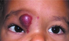
- Superficial lesions appear as a bright red, raised, lobulated (“strawberry”) mark on the eyelid skin
- Deeper lesions may show only a bluish discoloration through overlying normal-appearing skin, or may be entirely subcutaneous with no visible skin change at all, sometimes making the diagnosis less immediately obvious than for a classic superficial strawberry hemangioma
- The lesion characteristically enlarges with crying or straining (Valsalva-like maneuvers), from increased venous pressure transiently engorging the vascular lesion — a genuinely useful bedside sign supporting the diagnosis
- Size and growth rate are highly variable between patients, from small, cosmetically insignificant lesions to large masses causing substantial ptosis, globe displacement, or astigmatism
All-fit Slit-Lamp Adapter
Record and share exactly what you see at the slit lamp. One adapter fits any slit lamp or surgical microscope — and any smartphone.
From Choroida — the team behind this siteWhy Vision Is the Central Concern
A periocular capillary hemangioma large enough to physically occlude the visual axis, even partially, risks deprivation amblyopia, while a lesion pressing on the globe — even without any direct visual axis occlusion — can induce significant astigmatism and anisometropia, both independently amblyogenic.
Because the period of most rapid visual development in infancy is also, unfortunately, exactly when these lesions are undergoing their own most rapid growth, close monitoring and, when indicated, prompt treatment during this specific overlapping window is what actually protects vision, rather than waiting for the lesion’s natural involution, which occurs on a timescale far too slow to prevent amblyopia if the visual axis or corneal shape is significantly affected in the meantime.
Differential Diagnosis
- Orbital dermoid cyst — a distinct choristomatous lesion with its own characteristic firm, well-circumscribed quality and different natural history (stable rather than proliferative-then-involuting), discussed in its own dedicated article on this site
- Orbital lymphangioma — can also enlarge, sometimes acutely with intralesional hemorrhage, but generally does not show the same reliable spontaneous involution pattern as capillary hemangioma, discussed in its own dedicated article on this site
- Venous or lymphatic malformation more broadly — distinguished by imaging characteristics and, often, by a different growth pattern than the specific proliferation-then-involution trajectory of true infantile capillary hemangioma
- Rhabdomyosarcoma — a rare but important malignant mimic in a young child with a rapidly enlarging orbital mass, generally distinguished by a more aggressive growth rate and different imaging characteristics, and requiring prompt biopsy if there is genuine diagnostic uncertainty given the very different stakes involved
Diagnostic Evaluation
The diagnosis is often clinical, based on the characteristic appearance and growth pattern, though imaging (ultrasound or MRI) is useful for characterizing deeper lesions and confirming the diagnosis when the clinical picture is less certain.
Regular monitoring of refraction (looking for induced astigmatism), ocular alignment, and, most importantly, direct assessment of whether the lesion is encroaching on the visual axis is essential during the proliferative phase, given how quickly a rapidly growing lesion can begin to threaten vision.
Management
Small, non-vision-threatening lesions can be observed through their natural proliferative and involutional course, with regular monitoring to confirm they remain non-threatening to visual development.
Propranolol, a beta-blocker, has become first-line treatment for lesions requiring active intervention, and its introduction meaningfully changed the management of problematic infantile hemangiomas, because it is generally more effective and better tolerated than the corticosteroid treatment that was previously the mainstay. However, it requires appropriate initiation and monitoring given its systemic beta-blocking effects in an infant.
Intralesional or systemic corticosteroids remain an option, particularly when propranolol is contraindicated or insufficient.
Surgical excision is reserved for select cases: a well-localized lesion not responding adequately to medical therapy, or when rapid, definitive removal is needed to protect vision urgently.
Regardless of the specific treatment chosen, any associated amblyopia — from either direct visual axis occlusion or lesion-induced refractive error — needs its own dedicated treatment (patching or optical correction) alongside management of the hemangioma itself, because treating the tumor alone does not automatically reverse amblyopia that has already developed.
Families benefit from understanding from the outset that hemangioma treatment and amblyopia treatment are two separate, parallel tasks, since a shrinking lesion on its own does not guarantee that the child’s visual development is back on track without dedicated attention to the eye itself.


Document what you see
Two smartphone imaging tools built for everyday clinic use — one for the slit lamp, one for the fundus.
From Choroida — the team behind this siteReferences
- Léauté-Labrèze C, Hoeger P, Mazereeuw-Hautier J, et al. A randomized, controlled trial of oral propranolol in infantile hemangioma. New England Journal of Medicine.
- Schwartz SR, Blei F, Ceisler E, et al. Risk factors for amblyopia in children with capillary hemangiomas of the eyelids and orbit. Journal of AAPOS.
- American Academy of Ophthalmology. Basic and Clinical Science Course, Section 6: Pediatric Ophthalmology and Strabismus.
