
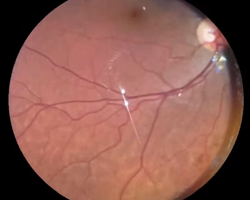
Vigabatrin retinal toxicity is a permanent, dose-related loss of peripheral vision caused by the antiepileptic drug vigabatrin.
The central acuity usually stays good, so patients often do not realize that their visual field has narrowed until the loss is advanced.
Because the drug controls seizures that are otherwise resistant, especially infantile spasms, the decision is rarely to avoid it altogether.
The ophthalmologist’s role is to establish a baseline, monitor for change, and help the neurologist weigh benefit against risk.
What Is Vigabatrin Retinal Toxicity?
Vigabatrin inhibits GABA transaminase, which raises brain GABA concentrations and suppresses seizures.
It is used for infantile spasms, particularly when caused by tuberous sclerosis complex, and for refractory focal seizures in adults.
Concentric visual field constriction was first reported in the 1990s in adults treated for epilepsy, and the association has since been confirmed in many series.
Retinal toxicity is thought to involve accumulation of GABA in the retina and damage to cone photoreceptors and inner retinal cells, although the mechanism is not fully understood.
The label carries a boxed warning, and in several countries vigabatrin is prescribed under a restricted-access safety program.
Who Is at Risk?
The prevalence of visual field defects among adults on long-term therapy is roughly a third or more in published series, and the range between studies is wide.
The main risk factors are:
- Higher cumulative dose and longer duration of therapy
- Male sex
- Older age in adults
- Preexisting visual field or retinal disease
- Concomitant treatment that may affect visual function, such as other antiepileptic drugs in some reports
Loss can appear within months in some patients and after years in others.
Children on short courses for infantile spasms have a lower risk than adults on long-term therapy, though toxicity has been documented in infants and young children.
Fundus Explorer Pro
Photograph the retinal findings described here with the phone already in your pocket — 22 D optics and built-in illumination in one handheld unit.
From Choroida — the team behind this siteClinical Features
Visual Fields
The characteristic defect is bilateral, symmetric concentric constriction, which begins in the nasal field and progresses.
Acuity is preserved until late, and color vision may be normal early on.
Patients may report bumping into objects, difficulty in crowds, or reduced peripheral awareness, although many are asymptomatic.
The damage is irreversible, and there is evidence of progression in some patients even after the drug is stopped.
Fundus
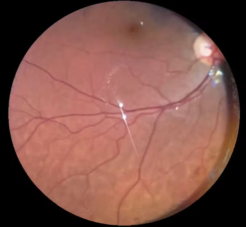
The fundus may look normal.
Some patients develop optic disc pallor, retinal vascular attenuation, or an abnormal macular reflex.
Absence of fundus change does not exclude toxicity.
OCT
Peripapillary RNFL thickness is reduced in many affected patients, and the loss correlates with the extent of visual field defect.
The thinning affects several sectors and tends to be more prominent nasally.
Macular OCT may show reduced retinal thickness in advanced cases.
OCT is objective and does not depend on patient cooperation, so it has become an important tool in children who cannot perform perimetry.
Electrophysiology
Full-field ERG shows reduced 30 Hz flicker amplitudes and delayed implicit times, reflecting cone dysfunction.
Photopic responses are affected earlier than scotopic ones, and the changes can precede visual field loss in some patients.
Electrophysiology is used in children and in patients who cannot perform reliable fields.
Differential Diagnosis
Concentric field loss has other causes.
- Glaucoma and other optic neuropathies (see optic atrophy)
- Retinitis pigmentosa and other rod-cone dystrophies
- Other drug toxicity, such as chloroquine and hydroxychloroquine (see hydroxychloroquine toxicity)
- Functional visual loss, which produces a tubular field that does not enlarge with distance
- Cortical and chiasmal disease
A history of vigabatrin exposure with a symmetric concentric defect and normal fundus is enough to raise the diagnosis.
Monitoring
Guidelines advise a baseline examination as soon as possible after starting treatment, ideally within a few weeks, and repeat testing at intervals of about every three to six months during therapy.
A practical schedule includes:
- Visual acuity and dilated fundus examination
- Automated perimetry with kinetic or static testing when the patient can cooperate
- OCT of the RNFL and macula
- ERG in young children and in patients who cannot do perimetry
- Testing after discontinuation, to document the final state
Reliability of testing is a problem in the very young and in patients with developmental disability, so combining several modalities gives more confidence.
Patients and caregivers should be told about the risk and about the need for regular testing, and consent should be documented.
Monitoring does not prevent damage, but it allows the neurologist to decide whether continuing is justified.
Management
There is no treatment that reverses the damage.
The decision to continue vigabatrin belongs to the neurologist together with the family, with the ophthalmologist supplying objective information.
Infantile Spasms
Vigabatrin is a first-line drug for infantile spasms, especially in tuberous sclerosis, and the benefit is substantial.
It is usually stopped once seizures have been controlled for a defined period, which limits cumulative exposure.
Adults
In adults, the drug is reserved for refractory epilepsy, and treatment should be stopped if no clear benefit occurs within the first months.
If field defects appear, the neurologist may reduce the dose or switch drugs when seizure control allows.
Supportive Care
Patients with established field loss benefit from low-vision assessment and counseling on driving, since visual field standards for driving licenses may not be met.
Prognosis
Field loss is permanent, and the extent depends on cumulative exposure.
Many patients with mild defects have normal daily function, and central vision and reading are usually spared.
Severe constriction limits mobility and driving.
Clinicians should document exposure carefully, since vigabatrin toxicity may be confused with other causes of field loss years later.


Document what you see
Two smartphone imaging tools built for everyday clinic use — one for the slit lamp, one for the fundus.
From Choroida — the team behind this siteReferences
- Eke T, Talbot JF, Lawden MC. Severe persistent visual field constriction associated with vigabatrin. BMJ. 1997;314:180-181.
- Kalviainen R, Nousiainen I. Visual field defects with vigabatrin: epidemiology and therapeutic implications. CNS Drugs. 2001;15:217-230.
- Wild JM, Ahn HS, Baulac M, et al. Vigabatrin and epilepsy: lessons learned. Epilepsia. 2007;48:1318-1327.
- Frisen L, Malmgren K. Characterization of vigabatrin-associated optic atrophy. Acta Ophthalmol Scand. 2003;81:466-473.
- Clayton LM, Devile M, Punte T, et al. Retinal nerve fiber layer thickness in vigabatrin-exposed patients. Ann Neurol. 2011;69:845-854.
- Westall CA, Wright T, Cortese F, Kumarappah A, Snead OC 3rd, Buncic JR. Vigabatrin retinal toxicity in children with infantile spasms: an observational cohort study. Neurology. 2014;83:2262-2268.
