
Case Study
A 56-year-old male presented for a routine eye examination without any visual complaints. His past medical history was unremarkable, and he had no history of ocular trauma or surgery.

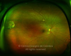
Fundoscopic examination of both eyes revealed multiple, well-demarcated, yellow-white patches in the peripheral retina, particularly in the inferior quadrant.
Optical coherence tomography (OCT) showed areas of retinal thinning corresponding to the lesions. Based on these findings, the patient was diagnosed with paving stone degeneration.
He was reassured about the benign nature of the condition and advised to return for regular follow-ups to monitor for any potential retinal complications, though none were anticipated.
Disease Entity
Paving stone degeneration, also known as cobblestone degeneration, is a benign peripheral retinal condition characterized by multiple, discrete, yellow-white lesions in the retinal periphery.
It is a common finding in older adults and is generally asymptomatic. Although it is associated with retinal atrophy, it does not significantly increase the risk of retinal detachment.
Fundus Explorer Pro
Photograph the retinal findings described here with the phone already in your pocket — 22 D optics and built-in illumination in one handheld unit.
From Choroida — the team behind this sitePathophysiology
Paving stone degeneration results from progressive chorioretinal atrophy, believed to be due to occlusion or drop out of the choriocapillaris. This leads to:
- Atrophy of the Retinal Pigment Epithelium (RPE): The RPE loses its function in affected areas, causing the underlying choroid to become visible as yellow-white lesions.
- Loss of Overlying Retina: The neurosensory retina above the atrophic RPE thins out but does not predispose to retinal tears.
- Formation of Pigment Clumps: Some cases may exhibit pigment migration around the atrophic patches, creating a mottled appearance.

Epidemiology
- Age: More common in individuals over 40 years of age.
- Sex: No clear gender predilection.
- Prevalence: Found in approximately 20–30% of normal adults during routine fundoscopic exams.
- Laterality: Frequently bilateral.
Clinical Features
Symptoms
- Typically asymptomatic.
- Rarely associated with flashes or floaters.
- No effect on central vision.
Fundoscopic Findings
- Small, round, or oval, yellow-white lesions in the retinal periphery.
- Lesions often appear in a clustered pattern, resembling “paving stones.”
- Commonly located in the inferior retinal periphery but can occur in other quadrants.
- The lesions may be surrounded by a pigmented border due to reactive RPE hyperplasia.
Diagnostic Evaluation
Paving stone degeneration is primarily diagnosed through ophthalmoscopic examination.
Fundus Photography
- Useful for documenting the extent and progression of lesions over time.
Optical Coherence Tomography (OCT)
- Shows retinal thinning over the areas of degeneration.
- May reveal loss of the RPE and outer retinal layers.
Fluorescein Angiography (FA)
- Not routinely required but may show window defects in areas of RPE atrophy.
Ultrawide-Field Imaging
- Helpful in visualizing peripheral retinal lesions.
Differential Diagnosis
Although paving stone degeneration is a distinct entity, it should be differentiated from other peripheral retinal conditions, such as:
- Retinal Lattice Degeneration: Thinner patches of the retina with a more networked appearance and higher risk of retinal tears.
- Retinal Holes: Often occur in lattice degeneration, whereas paving stone degeneration does not involve retinal breaks.
- Chorioretinal Scars: These may appear similar but are often associated with previous inflammation or infection.
- Peripheral Retinal Drusen: More scattered and associated with age-related changes.
Management
Observation
- No treatment is necessary in asymptomatic patients.
- Routine eye exams should be conducted to monitor for any peripheral retinal changes.
Patient Education
- Patients should be informed about the benign nature of paving stone degeneration.
- They should be aware of symptoms that might indicate retinal complications, such as flashes, floaters, or sudden vision changes, although these are uncommon.
Prognosis
- Excellent prognosis with no impact on vision.
- No known progression to retinal detachment or vision-threatening complications.
- Remains stable in most cases without requiring intervention.
Conclusion
Paving stone degeneration is a common, benign peripheral retinal condition characterized by atrophic patches of RPE and retina.
It is typically asymptomatic and requires no treatment, only routine monitoring. Differentiation from other peripheral retinal conditions is essential to reassure patients and avoid unnecessary interventions.


Document what you see
Two smartphone imaging tools built for everyday clinic use — one for the slit lamp, one for the fundus.
From Choroida — the team behind this siteReferences
- Straatsma BR, Foos RY, Heckenlively JR. Degenerative diseases of the peripheral retina. Ophthalmology. 1983;90(6):603-629.
- Wilkinson CP. Peripheral retinal degenerations and the risk of retinal detachment. Eye (Lond). 1986;10(1):57-65.
- Wang M, Sliney A, Lee K. Paving stone degeneration: a clinical review. Surv Ophthalmol. 2021;66(3):369-375.
- Chawla R, Dhasmana R, Azad S. Peripheral retinal changes: An overview. Indian J Ophthalmol. 2020;68(3):502-510.
- Byer NE. Long-term natural history of lattice degeneration of the retina. Ophthalmology. 1989;96(10):1396-1402.
Test yourself
A few questions straight from this article.
-
Paving stone degeneration is also known by which alternative name?
The article states paving stone degeneration is also called cobblestone degeneration, a benign peripheral retinal condition with discrete yellow-white lesions. -
What underlying vascular event is believed to cause paving stone degeneration?
The article attributes the progressive chorioretinal atrophy to occlusion or drop out of the choriocapillaris, which leads to RPE loss and thinning of the overlying retina. -
In what proportion of normal adults is paving stone degeneration found on routine fundoscopy?
The article gives a prevalence of roughly 20–30% of normal adults, most often over 40 years of age and frequently bilateral. -
Where in the retina are paving stone degeneration lesions most commonly located?
The article describes clustered yellow-white lesions most commonly in the inferior retinal periphery, although other quadrants may be involved. -
What does optical coherence tomography typically show in paving stone degeneration?
OCT demonstrates retinal thinning over the degenerative areas and may reveal loss of the RPE and outer retinal layers. -
Which feature distinguishes lattice degeneration from paving stone degeneration?
The article notes lattice degeneration shows thinner, networked retinal patches with a higher risk of retinal tears, whereas paving stone degeneration does not involve retinal breaks. -
Why may paving stone lesions be surrounded by a pigmented border?
The article attributes the pigmented rim around the atrophic patches to reactive hyperplasia of the retinal pigment epithelium. -
What is the appropriate management of asymptomatic paving stone degeneration?
No treatment is needed; the article advises observation with routine examinations plus patient education about the benign nature of the condition. -
What is the visual prognosis of paving stone degeneration?
The article describes an excellent prognosis: the lesions remain stable in most cases with no known progression to detachment or vision-threatening complications. -
Which imaging test best documents the extent of peripheral paving stone lesions over time?
The article recommends fundus photography for documenting lesion extent and progression, with ultrawide-field imaging helpful for visualising peripheral lesions.
