
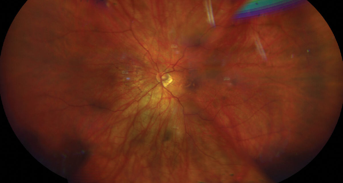
Pathologic myopia describes the structural complications of excessive axial elongation that damage the retina, choroid, and optic nerve and can cause irreversible vision loss.
High myopia and pathologic myopia are not the same thing, since many highly myopic eyes remain healthy for life.
The META-PM classification provides a common language for grading myopic maculopathy, and it predicts which eyes progress.
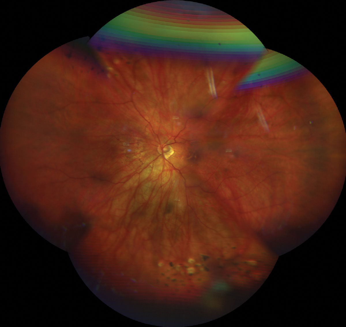
Defining Pathologic Myopia
High myopia is generally defined as a refractive error of -6.00 D or worse, or an axial length above 26 mm.
The International Myopia Institute defines pathologic myopia as the presence of a posterior staphyloma, myopic maculopathy at or beyond the diffuse atrophy stage, or myopic traction maculopathy and other complications attributable to axial elongation, in a highly myopic eye.
The distinction helps counseling, since pathologic myopia has a much higher risk of vision loss.
Risk is related to axial length, age, and the presence of staphyloma.
The prevalence of high myopia has been rising worldwide, and complications will follow in the coming decades (see also myopia control with red light and orthokeratology).
The META-PM Classification
The Meta-Analysis of Pathologic Myopia (META-PM) study group proposed a photographic classification of myopic maculopathy.
It uses five categories of fundus change and three additional lesions.
Categories
- C0: no myopic retinal lesions
- C1: tessellated fundus only
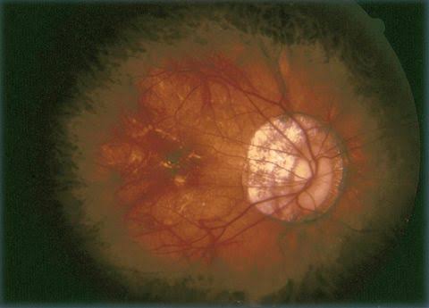
- C2: diffuse chorioretinal atrophy
- C3: patchy chorioretinal atrophy
- C4: macular atrophy
Plus Lesions
- Lacquer cracks, which are linear breaks in Bruch’s membrane
- Myopic choroidal neovascularization (see myopic CNV)
- Fuchs spot, a pigmented scar from an old neovascular membrane
Progression follows the categories.
Eyes at C2 or worse are at higher risk of developing patchy atrophy, and lacquer cracks are a risk factor for atrophy and myopic CNV.
Patchy atrophy tends to enlarge over the years, and it often threatens the fovea.
An updated ATN system grades atrophy, traction, and neovascularization separately, and it is useful for research and for describing complex eyes.
Fundus Explorer Pro
Photograph the retinal findings described here with the phone already in your pocket — 22 D optics and built-in illumination in one handheld unit.
From Choroida — the team behind this siteAssociated Complications
- Posterior staphyloma with a scleral outpouching that alters the macular contour (see staphyloma)
- Myopic traction maculopathy, including foveoschisis, foveal detachment, and macular hole (see myopic traction maculopathy)
- Macular hole retinal detachment in highly myopic eyes
- Myopic CNV, the most common cause of sudden central vision loss
- Dome-shaped macula (see dome-shaped macula)
- Peripheral retinal degeneration including lattice, with a higher risk of retinal tears and detachment (see lattice degeneration)
- Myopic optic neuropathy and glaucoma, with tilted discs and peripapillary atrophy
- Cataract, particularly nuclear and posterior subcapsular
A meta-analysis found that high myopia raises the risk of posterior subcapsular cataract, myopic maculopathy, retinal detachment, and open-angle glaucoma, and the risks increase with the degree of myopia.
Imaging
Widefield fundus photography or color imaging is used for META-PM grading.
OCT shows staphyloma, choroidal thinning, foveoschisis, and macular hole.
Swept-source OCT gives a better view of the choroid and sclera, and it can show dome-shaped macula and scleral changes.
OCT angiography helps detect myopic CNV without dye.
Fluorescein angiography confirms CNV, and ICGA may show choroidal vascular changes.
Autofluorescence shows atrophy.
Management
Monitoring
Patients with high myopia should be examined regularly, with attention to the macula and the peripheral retina.
They should be told to report new distortion, a central scotoma, floaters, or a curtain.
Amsler grid or home monitoring helps detect CNV.
Treatment of Complications
- Myopic CNV is treated with intravitreal anti-VEGF injections, based on the RADIANCE trial, which showed visual gains with ranibizumab
- Myopic traction maculopathy and macular hole may need vitrectomy with ILM peeling or macular buckle
- Retinal tears are treated with laser retinopexy, and retinal detachment needs surgical repair
- Glaucoma and cataract are managed in the usual way, with care regarding the thin sclera
There is no treatment that restores atrophic retina, and prevention focuses on slowing axial elongation in children.
Myopia Control in Children
Low-dose atropine, orthokeratology, multifocal soft contact lenses, spectacle lenses designed for myopia control, and outdoor time reduce the rate of axial elongation.
They lower the risk of high myopia in adulthood, and treatment decisions are individualized.
Low Vision and Support
Patients with irreversible loss benefit from low-vision assessment and rehabilitation.
Progression and Counseling
Myopic maculopathy progresses slowly and often silently.
Patients with tessellated fundus alone rarely progress to serious disease, whereas eyes with diffuse atrophy show a substantial chance of developing patchy atrophy over years.
Risk factors for progression include older age, longer axial length, the presence of staphyloma, and lacquer cracks.
Patients should understand several points:
- Their risk of retinal detachment is higher than average, and floaters or flashes need same-day evaluation
- Sudden central distortion may signal myopic CNV, and treatment works best when it is prompt
- Contact sports and activities with heavy trauma risk are worth discussing
- Refractive surgery and lens surgery do not remove the risk of retinal complications
- Their children should be examined early and offered myopia control
Regular follow-up every six to twelve months is reasonable for eyes with maculopathy, with imaging tailored to findings.
Prognosis
Visual loss develops slowly in most patients, and it often begins in the fifth and sixth decades.
Macular atrophy is the main cause of long-term visual impairment.
Regular examination and early treatment of CNV and traction can preserve vision.


Document what you see
Two smartphone imaging tools built for everyday clinic use — one for the slit lamp, one for the fundus.
From Choroida — the team behind this siteReferences
- Ohno-Matsui K, Kawasaki R, Jonas JB, et al. International photographic classification and grading system for myopic maculopathy. Am J Ophthalmol. 2015;159:877-883.
- Ohno-Matsui K, Wu PC, Yamashiro K, et al. IMI pathologic myopia. Invest Ophthalmol Vis Sci. 2021;62(5):5.
- Ruiz-Medrano J, Montero JA, Flores-Moreno I, Arias L, Garcia-Layana A, Ruiz-Moreno JM. Myopic maculopathy: current status and proposal for a new classification and grading system (ATN). Prog Retin Eye Res. 2019;69:80-115.
- Wolf S, Balciuniene VJ, Laganovska G, et al. RADIANCE: a randomized controlled study of ranibizumab in patients with choroidal neovascularization secondary to pathologic myopia. Ophthalmology. 2014;121:682-692.
- Haarman AEG, Enthoven CA, Tideman JWL, Tedja MS, Verhoeven VJM, Klaver CCW. The complications of myopia: a review and meta-analysis. Invest Ophthalmol Vis Sci. 2020;61(4):49.
- Baird PN, Saw SM, Lanca C, et al. Myopia. Nat Rev Dis Primers. 2020;6:99.
