
CASE REPORT
A 61–year–old man presented to consulting room with the complaint of metamorphopsia in the left eye. Visual acuity examination was performed on both eyes and yielded 8/10 -5.25 in the right eye and 5/10 -5.25 in the left eye.

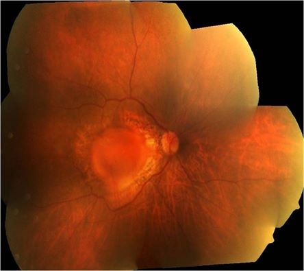
The Amsler test was found positive in the left eye. Anterior chamber examination was unremarkable for both eyes. Dilated fundus examination and fundus photography revealed vitreomacular traction outside the fovea and an epiretinal membrane (ERM) in the left eye.
SD-OCT (Cirrus HD-OCT, Carl Zeiss Meditec, Inc.) demonstrated partial detachment of the posterior hyaloid in the left eye with continued attachment at the fovea, abnormal foveal contour, and thickness, as well as an ERM.
SD-OCT is a relatively new optical coherence tomography device that allows three-dimensional structural imaging of the retina; its advantages over the classical time domain OCT, include precise point-by-point registration of the entire posterior pole of the eye and reproducibility of each scan.
With the use of SD-OCT, the precise location of pathology was identified and three-dimensional images of the macular area were obtained, revealing the vitreoretinal traction and the retinal folding as a result of the tangential traction of the ERM. Based on these SD-OCT findings, the diagnosis of myopic traction maculopathy was made.
Myopic Traction Maculopathy DISEASE entity
Myopic traction maculopathy (MTM), also known as myopic foveoschisis, is a schisis-like thickening of the retina in eyes with high myopia with posterior staphyloma. The pathologic features may also include lamellar or full-thickness macular holes, shallow foveal detachments, and inner retinal fluid.
It has been suggested that the schisis-like thickening represents edema from traction rather than a true schisis. MTM is estimated to affect between 9 and 34% of highly myopic eyes with posterior staphyloma. It is more prevalent in populations with high myopia and may be more prevalent in women.

In 1958, Phillips described localized posterior retinal detachments in patients with high myopia and posterior staphylomas without the presence of retinal holes. He postulated that myopic chorioretinal atrophy and the presence of a staphyloma were likely involved in the pathogenesis.
Prior to optical coherence tomography (OCT), MTM was difficult to distinguish from shallow retinal detachments and macular holes, particularly because slit-lamp biomicroscopy is limited in patients with myopic chorioretinal atrophy.
Takano and Kishi first reported the detection of MTM using OCT in 1999. They described foveal retinal detachment and foveoschisis in patients with high myopia with posterior staphylomas. Since then, spectral-domain OCT has been shown to be even more sensitive in detecting the pathologies associated with MTM.
Fundus Explorer Pro
Photograph the retinal findings described here with the phone already in your pocket — 22 D optics and built-in illumination in one handheld unit.
From Choroida — the team behind this siteMyopic Traction Maculopathy MANAGEMENT
The management of MTM is somewhat controversial because the natural course of the disease is not clear and the potential complications of surgical intervention are serious. Few small studies have looked at the natural disease course of MTM and found that some eyes remain stable for many years while other eyes progress to full-thickness macular holes or foveal detachments.
A recent larger study of MTM found that the incidence of progression to serious complications was related to the severity of macular retinoschisis. This correlation could be used to guide the timing and selection of patients for surgical intervention.
Premacular structures have also been reported to correlate to worse prognosis, and the presence of such structures could potentially be used to justify intervention. Some authors advocate that surgical treatment is indicated in MTM when VA is impaired or the patient complains of visual disturbances.
Another study showed persistent photoreceptor defects and irregular choroidal detachments in MFS patients with poor post-operative visual outcomes and recommends timing of surgery for MFS patients with there is threatened disruption of the outer retina on OCT.
PPV with ILM peel has been shown to result in the anatomic resolution of MTM and to improve visual acuity, even without the use of gas tamponade. ILM peeling relieves anterior traction on the macula and can remove scaffolding for cellular proliferation, possibly decreasing the risk of recurrence.
Removing the rigid ILM may also allow the retina to conform to the posterior staphyloma, which has been shown to contribute to macular detachment. One serious risk of ILM peeling in MTM is macular hole formation, which has been reported by several authors and observed to occur intraoperatively.
4 ILM peeling is technically difficult in patients with MTM because the ILM is thinner in highly myopic eyes, and may be more prone to tearing and iatrogenic macular hole formation. Interestingly, anatomic and visual acuity outcomes have been reportedly similar using PPV with and without ILM peel.
One option that addresses the need for an ILM peel with the risk of macular holes is an alternative fovea-sparing ILM peel. Various techniques have been described and limited studies have shown favorable outcomes. Scleral buckling has also been explored in the treatment of MTM.

The benefits of scleral buckling include avoiding cataracts and the need for cataract surgery, as well as avoiding iatrogenic macular holes associated with ILM peeling. Unlike PPV with ILM peeling, scleral buckling treats the structural contribution of the posterior staphyloma which may create shearing forces, stretching, and eventual failure of internal structures causing MTM.
Buckling also provides direct mechanical action to bring the RPE closer to the retina and may provide quicker resolution than a vitrectomy. The options for buckling include silicone bands or macular plombes, independently or in conjunction with PPV.
Suprachoroidal buckling has also been studied in a limited number of MTM patients and involves using a specially designed catheter, inserted into the suprachoroidal space, to inject hyaluronic acid into the area of the staphyloma to indent the choroid.
Though the majority of patients with scleral buckling in published studies had resolution of MTM and improved BCVA, complications included macular hole, recurrence of MTM, subretinal hemorrhage with and without choroidal neovascularization, choroidal detachment, RPE atrophy, and extrusion of the buckle.


Document what you see
Two smartphone imaging tools built for everyday clinic use — one for the slit lamp, one for the fundus.
From Choroida — the team behind this siteREFERENCES
- Panozzo G, Mercanti A. Optical coherence tomography findings in myopic traction maculopathy. Archives of ophthalmology. Oct 2004;122(10):1455-1460.
- Panozzo G, Mercanti A. Vitrectomy for myopic traction maculopathy. Archives of ophthalmology. Jun 2007;125(6):767-772.
- Baba T, Ohno-Matsui K, Futagami S, et al. Prevalence and characteristics of foveal retinal detachment without a macular hole in high myopia. American journal of ophthalmology. Mar 2003;135(3):338-342.
- Takano M, Kishi S. Foveal retinoschisis and retinal detachment in severely myopic eyes with posterior staphyloma. American journal of ophthalmology. Oct 1999;128(4):472-476.
- Phillips CI. Retinal detachment at the posterior pole. The British journal of ophthalmology. Dec 1958;42(12):749-753.
- Sayanagi K, Morimoto Y, Ikuno Y, Tano Y. Spectral-domain optical coherence tomographic findings in myopic foveoschisis. Retina. Apr 2010;30(4):623-628.
- Benhamou N, Massin P, Haouchine B, Erginay A, Gaudric A. Macular retinoschisis in highly myopic eyes. American journal of ophthalmology. Jun.
Test yourself
A few questions straight from this article.
-
Myopic traction maculopathy is also known by which other name?
The article defines myopic traction maculopathy (MTM), or myopic foveoschisis, as a schisis-like thickening of the retina in highly myopic eyes with posterior staphyloma. -
What proportion of highly myopic eyes with posterior staphyloma are estimated to have myopic traction maculopathy?
The article gives an estimated prevalence of 9 to 34% among highly myopic eyes with posterior staphyloma, and notes it may be more common in women. -
Which symptom did the patient in the myopic traction maculopathy case report describe?
The 61-year-old man presented with metamorphopsia of the left eye, and the Amsler test was positive on that side. -
Who first reported detection of myopic traction maculopathy using OCT, and in what year?
Takano and Kishi described foveal retinal detachment and foveoschisis on OCT in highly myopic eyes with posterior staphyloma in 1999. -
In 1958 Phillips described which finding in patients with high myopia and posterior staphyloma?
Phillips reported localized posterior retinal detachments in highly myopic staphylomatous eyes with no retinal holes, and implicated chorioretinal atrophy and the staphyloma in pathogenesis. -
Which advantage of SD-OCT over time-domain OCT is emphasised in this myopic traction maculopathy case?
The article credits SD-OCT with precise point-by-point registration of the entire posterior pole and reproducible scans, allowing three-dimensional imaging of the macula. -
In myopic traction maculopathy, progression to serious complications correlates best with which factor?
A larger study found the incidence of progression related to how severe the macular retinoschisis was, which can help guide timing and patient selection for surgery. -
How does internal limiting membrane peeling help in myopic traction maculopathy?
ILM peeling relieves anterior traction on the macula and removes the scaffold for cellular proliferation, possibly lowering recurrence risk. -
What is a recognised serious intraoperative risk of ILM peeling in myopic traction maculopathy?
Macular hole formation has been reported and observed intraoperatively; the ILM is thinner in highly myopic eyes and more prone to tearing. -
Suprachoroidal buckling for myopic traction maculopathy involves injecting which substance?
A specially designed catheter is passed into the suprachoroidal space to inject hyaluronic acid over the staphyloma, indenting the choroid.
