
Introduction
The worldwide prevalence of myopia is rising at an alarming rate, particularly among children and adolescents.
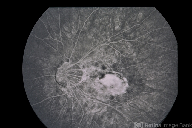
It is estimated that by 2050, nearly half of the global population will be affected by myopia, with significant public health implications.
High myopia increases the risk of sight-threatening complications such as retinal detachment, myopic macular degeneration, and glaucoma.
Thus, there is a growing emphasis on discovering and implementing effective strategies for myopia control.
Traditional methods, including atropine eye drops, orthokeratology lenses, and multifocal contact lenses, have demonstrated varying levels of success.
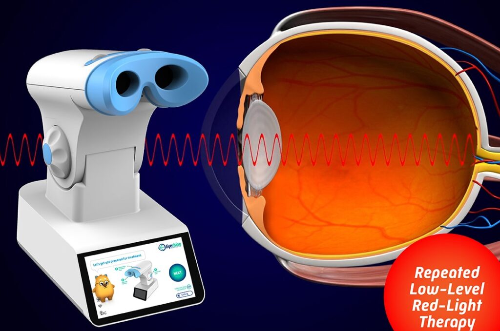
More recently, repeated low-level red light (RLRL) therapy has emerged as a promising non-invasive approach for myopia control.
While the mechanism behind RLRL’s efficacy is not yet fully understood, early studies have shown encouraging results in slowing axial elongation and myopic progression.
A novel aspect of investigation is whether RLRL therapy induces structural changes in the retina, particularly within the cone photoreceptor layer.
The integrity and density of cone cells are critical for central vision, color perception, and visual acuity. Any intervention that affects these cells warrants careful scrutiny.
Background
The human retina consists of millions of photoreceptors — rods for low-light vision and cones for color and fine detail.
In the macular region, particularly at the fovea, cone density peaks, facilitating high-resolution vision.
Myopia involves elongation of the eyeball, which can stretch and thin the retina, potentially leading to alterations in photoreceptor spacing and density.
Emerging therapies like RLRL have shown functional benefits, but their effects on retinal microstructure remain largely unexplored.
The potential impact on cone photoreceptors is of particular importance given their role in maintaining visual acuity and quality of life.
Fundus Explorer Pro
Photograph the retinal findings described here with the phone already in your pocket — 22 D optics and built-in illumination in one handheld unit.
From Choroida — the team behind this siteStudy Overview
Objective
The primary aim was to evaluate whether repeated low-level red light therapy induces measurable changes in cone density among myopic children.
Study Design
-
Type: Prospective, longitudinal cohort study.
-
Population: Children diagnosed with low to moderate myopia.
-
Intervention: Administration of RLRL therapy using a red light-emitting device.
-
Duration: Follow-up over several months.
-
Measurements: Quantitative analysis of cone density using high-resolution retinal imaging modalities.
Methods
Participants underwent baseline examinations, including cycloplegic refraction, axial length measurements, and adaptive optics imaging to capture detailed views of the cone mosaic.
The children then received RLRL therapy at specified frequencies and durations, consistent with previous protocols known to reduce myopia progression.
Post-treatment imaging was performed at regular intervals to monitor changes in cone density, spacing, and arrangement.
Results
Changes in Cone Density
-
Findings: After a course of RLRL therapy, significant changes were observed in the density and spatial organization of cone photoreceptors.
-
Magnitude: The cone density alterations were measurable but did not indicate widespread degeneration or loss.
-
Location: Changes were most prominent in the parafoveal region, where mechanical stress from ocular elongation may interact with treatment effects.
Functional Outcomes
Although the primary focus was on structural changes, the study also noted no significant decline in best-corrected visual acuity (BCVA), suggesting that cone function was preserved during the follow-up period.
Safety
No adverse events directly related to RLRL therapy were reported. The treatment was generally well tolerated, with high compliance rates among participants.
Discussion
Interpretation of Findings
The detection of cone density changes following RLRL therapy raises intriguing questions. Several hypotheses could explain these findings:
-
Mechanical Stretch Hypothesis: RLRL may alter scleral or choroidal biomechanics, indirectly affecting the retinal architecture and photoreceptor distribution.
-
Metabolic and Vascular Changes: Red light exposure has been proposed to enhance mitochondrial function and retinal blood flow, possibly promoting a microenvironment conducive to photoreceptor remodeling.
-
Photobiomodulation Effects: Low-level light therapy is known to modulate cellular activity, which may influence the maintenance, spacing, or turnover of cones.
Importantly, the absence of visual acuity deterioration suggests that the observed structural changes are not harmful in the short term. However, long-term implications remain unknown.
Comparison With Previous Studies
Earlier research primarily focused on axial length control and refractive outcomes following RLRL therapy. Few studies have addressed retinal microstructural changes.
This study pioneers the exploration of RLRL’s impact on the photoreceptor layer, adding a critical dimension to our understanding of myopia interventions.
Limitations
-
Short Follow-Up: Longer-term follow-up is needed to determine whether cone density stabilizes, continues to change, or reverses after therapy cessation.
-
Sample Size: Larger studies are necessary to validate these preliminary findings.
-
Functional Testing: Although BCVA remained stable, more sensitive functional tests such as microperimetry could reveal subtle visual changes not captured by standard acuity tests.
Clinical Implications
The findings have significant clinical ramifications:
-
Treatment Safety: Current evidence supports the short-term safety of RLRL therapy concerning retinal structure and function.
-
Monitoring Recommendations: Children undergoing RLRL therapy might benefit from regular retinal imaging to monitor potential structural changes.
-
Informed Consent: Physicians should discuss the possibility of retinal remodeling with parents and guardians when recommending RLRL therapy.
-
Future Directions: Deeper investigation into the biological mechanisms underlying these changes could lead to improved protocols optimizing both efficacy and safety.


Document what you see
Two smartphone imaging tools built for everyday clinic use — one for the slit lamp, one for the fundus.
From Choroida — the team behind this siteReferences
-
Li SM, Zhuang J, Xiong R, et al. Cone Density Changes After Red Light Treatment in Children With Myopia. JAMA Ophthalmology. 2024;142(5):491-499. doi:10.1001/jamaophthalmol.2024.1234
-
Morgan IG, Ohno-Matsui K, Saw SM. Myopia. Lancet. 2012;379(9827):1739-1748.
-
Hung LF, Arumugam B, She Z, et al. The effects of narrow-band long-wavelength lighting on refractive development in infant rhesus monkeys. Invest Ophthalmol Vis Sci. 2018;59(12):5668-5679.
-
Read SA, Vincent SJ, Collins MJ. Light exposure and eye growth in childhood. Invest Ophthalmol Vis Sci. 2015;56(11):6779-6787.
Test yourself
A few questions straight from this article.
-
What proportion of the global population is projected to be myopic by 2050?
Myopia prevalence is rising rapidly, and it is estimated that nearly half the global population will be affected by 2050, with major public health implications. -
Which sight-threatening complications are associated with high myopia?
High myopia raises the risk of retinal detachment, myopic macular degeneration and glaucoma, which is why effective myopia control strategies are being pursued. -
Repeated low-level red light therapy is used in children for which purpose?
RLRL therapy is a non-invasive approach to myopia control that has shown encouraging early results in slowing axial elongation and myopic progression. -
Which imaging technique was used to examine the cone mosaic in this study of red light therapy?
Baseline assessment included cycloplegic refraction, axial length measurement and adaptive optics imaging, which captures detailed views of the cone mosaic. -
Where were cone density changes most prominent after red light therapy in myopic children?
Changes in cone density and spatial organisation were most prominent parafoveally, where mechanical stress from ocular elongation may interact with treatment effects. -
What happened to best-corrected visual acuity during follow-up of red light therapy?
There was no significant decline in best-corrected visual acuity, suggesting cone function was preserved during the follow-up period despite the structural changes. -
Which mechanism was proposed to explain cone changes after red light therapy?
Proposed explanations include altered scleral or choroidal biomechanics, photobiomodulation effects, and enhanced mitochondrial function with increased retinal blood flow. -
What was reported about the safety and tolerability of red light therapy in this study?
No adverse events directly related to RLRL therapy were reported; the treatment was generally well tolerated with high compliance among participants. -
Which test was suggested to detect subtle visual changes missed by standard acuity testing?
Although BCVA remained stable, the authors note that more sensitive functional tests such as microperimetry could reveal subtle changes standard acuity does not capture. -
What monitoring is recommended for children receiving repeated low-level red light therapy?
Children undergoing RLRL therapy may benefit from regular retinal imaging to monitor potential structural changes, and parents should be counselled about possible retinal remodelling.
