
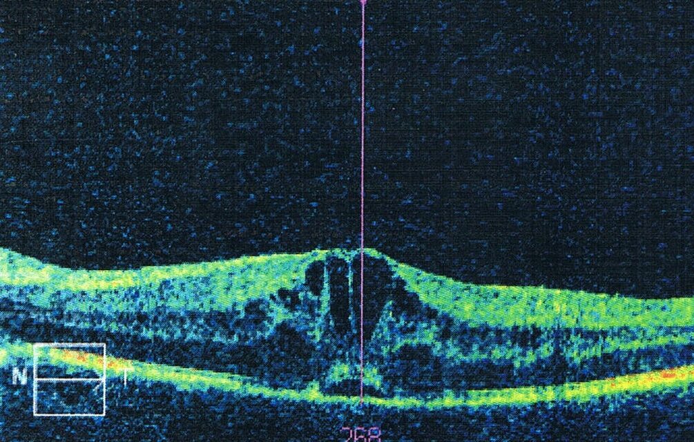
Uveitic macular edema (UME) is fluid accumulation in the macula caused by intraocular inflammation, and it is the most common structural cause of visual loss in patients with uveitis.
It may accompany any anatomical form of uveitis, but it is especially frequent in intermediate, posterior, and panuveitis.
The edema is a consequence of inflammation, so treatment that targets only the fluid usually fails when the underlying disease stays active.
This article reviews how UME develops, how to identify it on imaging, and how to escalate treatment when the first step is not enough.
Pathogenesis of Uveitic Macular Edema
Inflammatory cytokines such as IL-6, TNF-alpha, and VEGF disrupt the tight junctions of the retinal capillary endothelium and the retinal pigment epithelium.
This breaks down the inner and outer blood-retinal barriers, and fluid leaks into the retina faster than it can be removed.
The RPE pump, which normally clears subretinal fluid, may also be impaired by inflammation.
Chronic edema damages Müller cells and photoreceptors, and the visual loss then becomes irreversible.
Vitreoretinal traction and epiretinal membranes are common in uveitic eyes and add a mechanical component.
In a large series of patients with intraocular inflammation, macular edema was among the leading causes of visual impairment.
Clinical Presentation
Patients notice blurred central vision, metamorphopsia, or reduced contrast.
Many have symptoms of the underlying uveitis, such as floaters, redness, or photophobia.
Vitreous haze may obscure the macula on examination, so imaging is often needed to see the edema.
UME can be the first sign of uveitis, particularly in intermediate uveitis and birdshot chorioretinopathy.
Fundus Explorer Pro
Photograph the retinal findings described here with the phone already in your pocket — 22 D optics and built-in illumination in one handheld unit.
From Choroida — the team behind this siteImaging
OCT
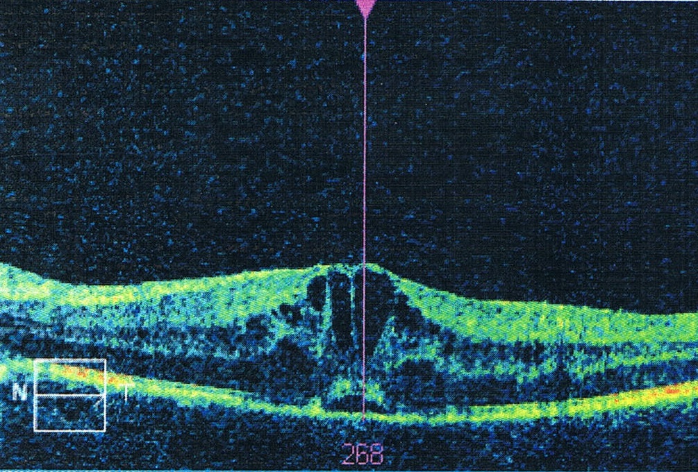
OCT is the standard tool for diagnosis and monitoring.
Typical patterns are:
- Cystoid spaces in the inner and outer retinal layers
- Diffuse retinal thickening without distinct cysts
- Subretinal fluid, which is more common in uveitic edema than in diabetic edema and often accompanies serous detachment
- Epiretinal membrane or vitreomacular traction
- Disruption of the ellipsoid zone and external limiting membrane in chronic disease
Central subfield thickness is used to follow treatment, and it correlates only moderately with visual acuity.
Fluorescein Angiography
Angiography shows petaloid leakage in the macula and often disc hyperfluorescence.
It also identifies retinal vasculitis, capillary nonperfusion, and peripheral leakage that guide systemic treatment.
Differential Diagnosis
Other causes of macular edema in a patient with uveitis should be considered:
- Pseudophakic cystoid macular edema, since many uveitic eyes have had cataract surgery (see cystoid macular edema)
- Diabetic macular edema
- Retinal vein occlusion
- Hypotony maculopathy, which may follow ciliary body shutdown
- Steroid-related complications such as central serous chorioretinopathy
- Drug toxicity
Identifying a second cause matters because treatment differs.
Management
Control the Underlying Uveitis
Treatment starts with controlling the inflammation.
This may involve corticosteroids and steroid-sparing immunosuppression such as methotrexate, mycophenolate, azathioprine, or biologics such as adalimumab.
Infectious causes need specific therapy first.
Many cases of UME improve as the systemic disease comes under control.
Local Corticosteroids
Local steroids act quickly and are the usual first step for unilateral or asymmetric edema.
- Periocular triamcinolone given by the posterior sub-Tenon’s route is well tolerated and has a lower risk of raised IOP
- Intravitreal triamcinolone is more potent and has a shorter duration of action
- Dexamethasone implant (0.7 mg) releases drug for several months, and in the HURON trial it improved vitreous haze in nearly half of treated eyes compared with about one in eight in the sham group
- Fluocinolone acetonide implants offer long-term control, and they cause cataract in nearly all phakic eyes and raised IOP in a substantial proportion
The POINT trial compared periocular triamcinolone, intravitreal triamcinolone, and the dexamethasone implant in uveitic macular edema.
All three reduced central subfield thickness, and the intravitreal treatments produced a larger reduction than periocular triamcinolone, at the cost of a higher risk of elevated IOP.
Patients with a history of steroid response, glaucoma, or ocular hypertension need close IOP monitoring after any steroid injection.
Systemic Corticosteroids
Oral prednisone or intravenous methylprednisolone control severe or bilateral disease, and a short course is often used to start treatment.
They are not suitable for long-term use because of side effects.
Other Options
- Carbonic anhydrase inhibitors such as acetazolamide can reduce edema in some patients, although the effect is often modest
- Interferon alpha has been used for resistant edema, especially in Behçet disease
- Anti-VEGF injections are less effective than in diabetic edema, but they may help when VEGF-driven leakage is prominent
- Topical NSAIDs have limited benefit in most cases
- Intravitreal methotrexate is used in some centers for eyes that cannot tolerate steroids
Surgery
Vitrectomy with membrane peeling helps when there is a taut epiretinal membrane or vitreomacular traction.
It can also improve inflammatory control in some eyes, though evidence is limited.
Special Situations
Juvenile Idiopathic Arthritis and Pediatric Uveitis
Children develop macular edema less often, but it can occur in chronic uveitis, and it is easy to overlook because young patients do not report symptoms.
Periocular steroids and implants carry a higher risk of cataract and glaucoma in this group, and early biologic therapy is often preferred (see JIA-associated uveitis).
Birdshot Chorioretinopathy and Sarcoid Uveitis
In birdshot disease, macular edema is a leading cause of central vision loss, and it may persist after the vitritis is controlled (see birdshot chorioretinopathy).
Sarcoid uveitis produces chronic edema that responds to systemic therapy and to local steroids (see sarcoid uveitis).
After Cataract Surgery
Eyes with uveitis are at high risk of cystoid edema after cataract surgery.
Surgery should be performed when inflammation has been quiet for at least three months, with perioperative steroid cover.
A Practical Stepwise Approach
A reasonable sequence for a patient with newly detected edema is as follows.
- Confirm active inflammation, and exclude infection before increasing immunosuppression
- Start or optimize systemic therapy if disease is bilateral, chronic, or associated with systemic disease
- Add a local steroid for the edematous eye, choosing the route according to laterality, lens status, and IOP history
- Reassess with OCT at four to eight weeks
- Switch route or agent if the response is partial, and consider vitrectomy if there is a mechanical cause
- Review IOP after every steroid injection and at regular intervals
The endpoint is a dry macula with no active inflammation on the lowest effective treatment.
Prognosis and Follow-Up
Edema that resolves quickly usually leaves acuity intact.
Chronic edema causes photoreceptor loss, and eyes that have had edema for many months may not regain vision even after the retina dries.
Recurrence follows tapering of inflammatory control, and repeated OCT is the best way to detect it early.
Management of UME is shared between the retina specialist and the uveitis physician, and success usually requires control of the disease and of the fluid.


Document what you see
Two smartphone imaging tools built for everyday clinic use — one for the slit lamp, one for the fundus.
From Choroida — the team behind this siteReferences
- Rothova A, Suttorp-van Schulten MS, Frits Treffers W, Kijlstra A. Causes and frequency of blindness in patients with intraocular inflammatory disease. Br J Ophthalmol. 1996;80:332-336.
- Lowder C, Belfort R Jr, Lightman S, et al. Dexamethasone intravitreal implant for noninfectious intermediate or posterior uveitis. Arch Ophthalmol. 2011;129:545-553.
- Thorne JE, Sugar EA, Holbrook JT, et al. Periocular triamcinolone vs. intravitreal triamcinolone vs. intravitreal dexamethasone implant for the treatment of uveitic macular edema: the POINT trial. Ophthalmology. 2019;126:283-295.
- Kempen JH, Altaweel MM, Holbrook JT, et al. Randomized comparison of systemic anti-inflammatory therapy versus fluocinolone acetonide implant for intermediate, posterior, and panuveitis: the Multicenter Uveitis Steroid Treatment Trial. Ophthalmology. 2011;118:1916-1926.
- Jaffe GJ, Dick AD, Brezin AP, et al. Adalimumab in patients with active noninfectious uveitis. N Engl J Med. 2016;375:932-943.
