
Retinal hemorrhage takes different shapes depending on the layer where the blood collects, and the shape gives a first clue to the cause.
A flame-shaped hemorrhage in the nerve fiber layer, a round dot in the inner nuclear layer, and a boat-shaped collection in front of the retina reflect different anatomy and different diseases.
Learning to name the layer before naming the disease makes fundus examination more systematic and helps with the next test to order.
Why the Shape of a Retinal Hemorrhage Matters
Blood spreads along the planes of least resistance.
In the nerve fiber layer the axons run parallel to the surface, so blood forms linear or flame-shaped streaks that follow the fibers.
In the deeper layers, the vertical orientation of the cells and the compact structure restrict spread, and blood forms round or blot-like collections.
Blood in front of the retina collects in the potential spaces beneath the internal limiting membrane or the posterior hyaloid.
Blood beneath the retina or the RPE is dark and lies beneath the retinal vessels.
The location gives a clue about the source vessel.
Types of Retinal Hemorrhage
Flame-Shaped (Nerve Fiber Layer)
- Feathery, red streaks that follow the arcuate nerve fiber pattern, often near the disc
- Arise from superficial radial peripapillary capillaries
- Seen in hypertensive retinopathy, retinal vein occlusion, papilledema, and disc swelling from other causes (see hypertensive retinopathy)
- Seen at the edge of a swollen disc in papilledema, and in vein occlusions of the affected sector
Dot and Blot (Deep Retinal)
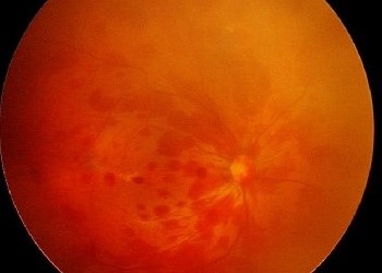
- Round, sharply demarcated dots and blots in the inner nuclear and outer plexiform layers
- Arise from the deep capillary plexus and microaneurysms
- Typical of diabetic retinopathy, and also seen in vein occlusion, ocular ischemic syndrome, and hypertension
- Blot hemorrhages are larger, and clusters suggest ischemia
Preretinal or Subhyaloid
- Boat-shaped or crescent hemorrhage with a flat upper border, sometimes with a fluid level
- Collects between the ILM and the posterior hyaloid
- Seen in proliferative diabetic retinopathy, Valsalva retinopathy, Terson syndrome, and trauma (see Valsalva retinopathy and Terson syndrome)
Sub-ILM
- Smooth, dome-shaped collection that does not layer
- Seen in Valsalva retinopathy and trauma
Subretinal
- Dark red or gray-green, deeper than the retinal vessels, with a smooth margin
- Seen in neovascular AMD, retinal macroaneurysm, trauma, and choroidal tumors (see macroaneurysm)
Sub-RPE
- Dark, well-demarcated dome, sometimes brown-black
- Seen in polypoidal choroidal vasculopathy and choroidal neovascularization
- Can mimic choroidal melanoma
Roth Spots
- Hemorrhages with a white or pale center
- Historically linked to bacterial endocarditis, but also seen in leukemia, anemia, diabetes, hypoxia, and connective tissue disease (see Roth spots)
- The white center may be a fibrin-platelet plug or a cluster of inflammatory or neoplastic cells
Vitreous Hemorrhage
Blood in the vitreous gives haze or a dense obscuration of the fundus (see vitreous hemorrhage).
Fundus Explorer Pro
Photograph the retinal findings described here with the phone already in your pocket — 22 D optics and built-in illumination in one handheld unit.
From Choroida — the team behind this siteDistribution Patterns
The distribution across the fundus helps as much as the layer.
- Diffuse in all quadrants: central retinal vein occlusion, hyperviscosity, severe anemia or thrombocytopenia (see central retinal vein occlusion)
- Sectoral: branch retinal vein occlusion, following the territory of the affected vein (see branch retinal vein occlusion)
- Peripapillary: papilledema, disc swelling, disc hemorrhage in glaucoma
- Macular: Valsalva retinopathy, macroaneurysm, trauma, neovascular AMD
- Midperipheral, mostly deep: ocular ischemic syndrome and diabetic retinopathy
- Multiple layers, extending to the periphery: abusive head trauma, a pattern that needs careful documentation (see shaken baby syndrome)
- Bilateral and symmetric: systemic disease such as hypertension, blood disorders, or diabetes
Systemic Causes to Consider
- Diabetes and hypertension
- Blood disorders: anemia, thrombocytopenia, leukemia, hyperviscosity
- Anticoagulant and antiplatelet therapy
- Infective endocarditis and sepsis
- Vasculitis and connective tissue disease
- Increased venous pressure from straining, vomiting, or chest compression (Purtscher-like patterns are described in Purtscher retinopathy)
- High-altitude exposure
- Head and chest trauma
- Neonatal hemorrhages after vaginal delivery, which are common and usually resolve within weeks
A blood pressure check, glucose, and complete blood count are the minimum initial tests.
Investigations
- Dilated fundus examination with scleral depression, since peripheral findings help distinguish causes
- Fundus photographs, ideally widefield, for documentation
- OCT to place the hemorrhage in a layer, and to identify macular edema
- Fluorescein angiography to show the source of bleeding, leakage, and nonperfusion, recognizing that blood blocks fluorescence
- B-scan ultrasonography when the view is obscured
- Systemic tests directed by the pattern
Mimics and Pitfalls
Several findings are mistaken for retinal hemorrhage.
- Pigmented lesions such as a choroidal nevus, CHRPE, or a melanocytoma, which do not change with time and lack the feathery margins of blood
- Vascular tufts and macroaneurysms, which are the source of the bleed and may be hidden by it
- Retinal neovascularization, whose fronds may be surrounded by hemorrhage
- Sub-RPE blood over a polypoidal lesion, which may look like a melanoma
Dates matter as well.
Fresh blood is bright red, and it turns yellow-brown as it ages, so a change in color over days to weeks supports hemorrhage.
A lesion that stays dark and unchanged for months should be reassessed for a pigmented tumor.
Management
Management addresses the cause.
- Hypertension and diabetes require systemic control
- Retinal vein occlusion is treated for macular edema and neovascularization
- Bleeding disorders and drug effects need medical management
- Preretinal hemorrhage may be observed or treated with laser hyaloidotomy or vitrectomy if it blocks central vision
- Trauma needs a full examination for other injuries
- Suspected abusive head trauma needs urgent multidisciplinary evaluation according to local child protection protocols
Most hemorrhages resolve over weeks, and the visual outcome depends on the damage to the underlying retina.


Document what you see
Two smartphone imaging tools built for everyday clinic use — one for the slit lamp, one for the fundus.
From Choroida — the team behind this siteReferences
- Schachat AP, Sadda SR, Hinton DR, Wilkinson CP, Wiedemann P, eds. Ryan’s Retina. 6th ed. Philadelphia: Elsevier; 2018.
- Salmon JF. Kanski’s Clinical Ophthalmology: A Systematic Approach. 9th ed. Philadelphia: Elsevier; 2020.
- Levin AV, Christian CW; Committee on Child Abuse and Neglect, Section on Ophthalmology. The eye examination in the evaluation of child abuse. Pediatrics. 2010;126:376-380.
- Emerson MV, Pieramici DJ, Stoessel KM, Berreen JP, Gariano RF. Incidence and rate of disappearance of retinal hemorrhage in newborns. Ophthalmology. 2001;108:36-39.
- Wong TY, Mitchell P. Hypertensive retinopathy. N Engl J Med. 2004;351:2310-2317.
Test yourself
A few questions straight from this article.
-
Why does a nerve fibre layer retinal haemorrhage take a flame shape?
Blood spreads along planes of least resistance, and in the nerve fibre layer the axons run parallel to the retinal surface, so haemorrhage forms linear or flame-shaped streaks following the fibres. -
Which vessels give rise to flame-shaped haemorrhages near the optic disc?
Flame-shaped haemorrhages arise from the superficial radial peripapillary capillaries, which is why they appear near the disc in hypertensive retinopathy, vein occlusion and papilloedema. -
From which source do dot and blot retinal haemorrhages arise?
Dot and blot haemorrhages lie in the inner nuclear and outer plexiform layers, arising from the deep capillary plexus and microaneurysms, and clusters of blots suggest ischaemia. -
A boat-shaped haemorrhage with a flat upper border and a fluid level lies in which plane?
Preretinal or subhyaloid blood collects between the internal limiting membrane and the posterior hyaloid, forming a boat-shaped or crescent haemorrhage with a flat upper border. -
Which appearance identifies a haemorrhage as subretinal rather than intraretinal?
Subretinal blood appears dark red or grey-green with a smooth margin and lies deeper than the retinal vessels; it is seen in neovascular AMD, macroaneurysm, trauma and choroidal tumours. -
A dark, well-demarcated brown-black dome under the retinal pigment epithelium can be mistaken for which lesion?
Sub-RPE haemorrhage, seen with polypoidal choroidal vasculopathy and choroidal neovascularisation, forms a dark well-demarcated dome that can mimic choroidal melanoma. -
What may account for the pale centre of a Roth spot?
The white centre may be a fibrin-platelet plug or a cluster of inflammatory or neoplastic cells; although historically linked to bacterial endocarditis, Roth spots also occur in leukaemia, anaemia, diabetes and hypoxia. -
Retinal haemorrhages spread diffusely through all four quadrants suggest which group of causes?
A diffuse haemorrhage pattern in all quadrants points to central retinal vein occlusion, hyperviscosity, or severe anaemia or thrombocytopenia, whereas a sectoral pattern follows a branch vein territory. -
A dark fundus lesion has stayed unchanged in colour for several months. What does this suggest?
Fresh blood is bright red and turns yellow-brown as it ages, so a lesion that stays dark and unchanged for months should be reassessed as a pigmented tumour. -
How is a preretinal haemorrhage that blocks central vision managed?
Preretinal haemorrhage may simply be observed, but laser hyaloidotomy or vitrectomy is considered when it blocks central vision; most haemorrhages resolve over weeks.

Writes and reviews the clinical articles on Educate. Every post is checked against peer-reviewed sources before it goes live.


