
An infant who cries in bright light, waters constantly and screws their eyes shut is often assumed to have a blocked tear duct. Sometimes the pressure inside the eye is destroying the optic nerve.
Primary congenital glaucoma is raised intraocular pressure from a developmental abnormality of the aqueous drainage angle, present from birth or the first years of life.
An infant eye is elastic, so instead of simply cupping the disc, the whole globe stretches.
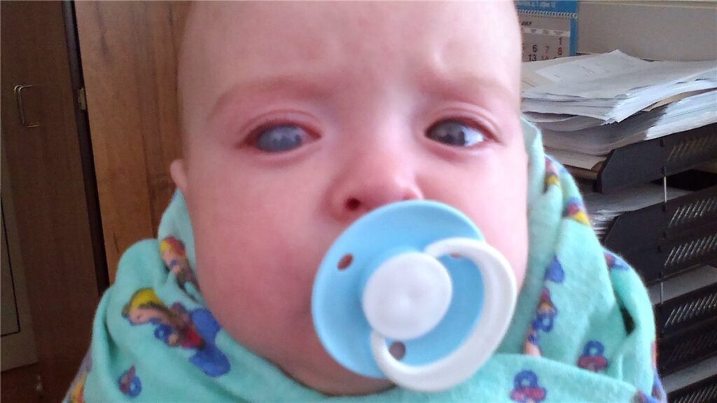
That stretching produces the signs that make this diagnosis possible from the end of the bed – an enlarged, cloudy cornea in a photophobic, watering baby.
Unlike adult glaucoma, this is a surgical disease from the outset; drops are a holding measure, not a treatment.
Recognising the classic triad and referring the same day is what preserves vision in these children.
What Is Primary Congenital Glaucoma?
Primary congenital glaucoma (PCG) is glaucoma resulting from isolated maldevelopment of the trabecular meshwork and anterior chamber angle, without other ocular or systemic anomalies.
It is classified by age at presentation:
- Newborn or true congenital – present at birth, roughly a quarter of cases
- Infantile – presenting between one month and two years, the largest group
- Late-recognised or juvenile-onset – presenting after two years, when the globe is less elastic and enlargement is less prominent
The word “primary” is doing important work here – secondary childhood glaucomas from aniridia, Axenfeld-Rieger anomaly, Sturge-Weber syndrome or previous cataract surgery are managed differently and must be excluded.
Epidemiology
PCG is rare in absolute terms but is a leading cause of childhood blindness where it goes unrecognised.
- The majority of cases present within the first year of life
- Bilateral involvement occurs in most cases, though frequently asymmetric
- Boys are affected more often than girls in most reported series
- Incidence is considerably higher in populations with high rates of consanguineous marriage
- Most cases are sporadic, but an autosomal recessive pattern with variable penetrance is well recognised, and CYP1B1 mutations are the most frequently implicated
Asymmetry is a diagnostic trap: a mildly affected fellow eye can look normal by comparison, so both eyes need measuring rather than comparing.
All-fit Slit-Lamp Adapter
Record and share exactly what you see at the slit lamp. One adapter fits any slit lamp or surgical microscope — and any smartphone.
From Choroida — the team behind this sitePathophysiology
The disease is a failure of normal angle development, and the infant eye’s response to pressure is what shapes the clinical picture.
- Maldevelopment of the trabecular meshwork and angle structures obstructs aqueous outflow
- Intraocular pressure rises and, because the infant sclera and cornea are elastic, the whole globe enlarges – buphthalmos
- Corneal enlargement stretches Descemet membrane until it splits, producing horizontal or curvilinear Haab striae
- Breaks in Descemet membrane allow aqueous into the stroma, causing corneal oedema, haze and the photophobia and watering that dominate the presentation
- Axial elongation produces progressive myopia and anisometropia
- Sustained pressure damages the optic nerve, and infant discs can cup rapidly – though this cupping is partly reversible if pressure is controlled early
The reversibility of infant disc cupping is one of the genuinely encouraging features of this disease, and it is a direct argument for treating quickly.
Clinical Presentation
The Classic Triad
- Epiphora – persistent watering
- Photophobia – distress and eye closure in bright light
- Blepharospasm – forced lid closure
These three symptoms in an infant should trigger a pressure check, not a diagnosis of blocked tear duct.
Examination Findings
- Buphthalmos – a visibly enlarged eye, often asymmetric between the two sides
- Corneal enlargement, with a horizontal diameter above roughly 12 mm in the first year being suspicious
- Corneal oedema and haze, which can range from subtle to a completely opaque cornea
- Haab striae – horizontal breaks in Descemet membrane, a highly specific sign
- Raised intraocular pressure, remembering that general anaesthetic agents lower measured pressure
- Optic disc cupping, often large and sometimes asymmetric
- Deep anterior chamber and, on gonioscopy, a high, flat iris insertion
Haab striae are worth looking for specifically, because they distinguish glaucomatous corneal enlargement from other causes of a cloudy neonatal cornea.
Diagnostic Evaluation
Examination Under Anaesthesia
- Most infants require examination under anaesthesia for reliable assessment
- Intraocular pressure measurement, interpreted with the knowledge that most anaesthetic agents reduce it and may mask disease
- Corneal diameter measurement in both eyes with callipers
- Gonioscopy to assess angle structures and exclude secondary causes
- Optic disc assessment and, where possible, photography for later comparison
Supporting Measurements
- Axial length by ultrasound biometry – progressive elongation is a sensitive marker of ongoing pressure damage and of treatment failure
- Cycloplegic refraction, which typically shows progressive myopia
- Pachymetry, since corneal thickness influences pressure readings
Excluding Secondary Glaucoma
- Careful examination for aniridia, Axenfeld-Rieger anomaly, Peters anomaly and the facial naevus of Sturge-Weber syndrome
- Systemic and genetic assessment where a syndromic association is suspected
Serial axial length is one of the most useful follow-up measurements available in infants, because it tracks disease control even when pressure readings are unreliable.
Differential Diagnosis
Conditions that can mimic the presentation include:
- Congenital nasolacrimal duct obstruction – watering and discharge, but no photophobia, normal corneal size and a clear cornea
- Congenital corneal opacities – Peters anomaly, sclerocornea, congenital hereditary endothelial dystrophy
- Birth trauma with forceps-related Descemet breaks – typically vertical or oblique, unlike the horizontal striae of glaucoma
- Megalocornea – a large but clear cornea with normal pressure and no disc changes
- Congenital rubella and metabolic causes of corneal clouding
- High myopia with a large eye but normal pressure
The orientation of Descemet breaks is a genuinely useful discriminator: horizontal or curvilinear suggests glaucoma, vertical suggests forceps injury.
Management
Surgery Is Definitive
- Angle surgery is first-line: goniotomy where the cornea is clear enough to visualise the angle, or trabeculotomy where it is not
- Success rates for angle surgery in classic PCG are good, and the procedure may be repeated
- Trabeculectomy with antimetabolite, or a glaucoma drainage device, for cases failing angle surgery
- Cyclodestructive procedures are generally reserved for refractory disease or eyes with poor visual potential
Medical Therapy as a Bridge
- Topical agents are used to lower pressure temporarily before surgery or to supplement incomplete surgical control
- Beta-blockers require caution in infants because of the risk of apnoea and bradycardia
- Brimonidine is contraindicated in infants and young children owing to the risk of CNS depression and apnoea
- Carbonic anhydrase inhibitors, topical or systemic, are commonly used
Visual Rehabilitation
- Correction of the myopia and anisometropia that follow axial elongation
- Amblyopia therapy, which is frequently the limiting factor in the final visual result
- Lifelong follow-up, since pressure can rise again years after apparently successful surgery
Even a perfectly controlled pressure produces a poor visual outcome if the resulting anisometropic amblyopia is not treated with equal commitment.
Prognosis
Outcomes have improved considerably with early angle surgery, but depend heavily on timing.
- Children presenting between about three and twelve months with clear corneas generally have the best surgical prognosis
- Presentation at birth with severe corneal clouding carries a poorer outlook
- Optic disc cupping can partially reverse in infants once pressure is controlled, unlike in adults
- Amblyopia from corneal scarring, anisometropia and induced myopia is a major cause of persistent visual loss even after successful pressure control
- Lifelong monitoring is essential, as late pressure rise and progressive damage can occur years later
The recurring theme is that pressure control is necessary but not sufficient – the visual result is decided by refraction and amblyopia therapy over the following years.


Document what you see
Two smartphone imaging tools built for everyday clinic use — one for the slit lamp, one for the fundus.
From Choroida — the team behind this siteReferences
- Childhood Glaucoma Research Network. Classification of childhood glaucoma. Consensus series.
- Yu Chan JY, Choy BNK, Ng ALK, Shum JWH. Review on the management of primary congenital glaucoma. Journal of Current Glaucoma Practice. 2015.
- Ho CL, Walton DS. Primary congenital glaucoma: 2004 update. Journal of Pediatric Ophthalmology and Strabismus. 2004.
- Primary Congenital Glaucoma. StatPearls, NCBI Bookshelf.
- Primary Congenital Glaucoma. EyeWiki, American Academy of Ophthalmology.
Test yourself
A few questions straight from this article.
-
What underlies primary congenital glaucoma?
Primary congenital glaucoma results from isolated maldevelopment of the trabecular meshwork and anterior chamber angle, without other ocular or systemic anomalies. -
Which triad of infant symptoms should prompt an intraocular pressure check rather than reassurance?
Persistent watering, distress in bright light and forced lid closure are the classic triad, and they should trigger a pressure check rather than a diagnosis of blocked tear duct. -
Why does raised pressure in an infant eye produce buphthalmos rather than isolated disc cupping?
Because the infant sclera and cornea are elastic, sustained pressure enlarges the whole globe, producing buphthalmos alongside optic nerve damage. -
What are Haab striae in primary congenital glaucoma?
Corneal enlargement stretches Descemet membrane until it splits, producing horizontal or curvilinear Haab striae, a highly specific sign that lets aqueous into the stroma. -
Above roughly what horizontal corneal diameter is an infant cornea in the first year suspicious for glaucoma?
A horizontal corneal diameter above roughly 12 mm in the first year of life is suspicious, and both corneas should be measured with callipers rather than compared by eye. -
How do forceps-related Descemet breaks differ from those of congenital glaucoma?
The orientation of the breaks discriminates the two: horizontal or curvilinear striae suggest glaucoma, while vertical or oblique breaks suggest forceps injury at birth. -
What caveat applies to intraocular pressure measured under general anaesthesia in an infant?
Most anaesthetic agents reduce measured intraocular pressure, so a normal reading under anaesthesia can mask genuine disease and must be read alongside the other signs. -
What is the first-line surgical treatment for primary congenital glaucoma?
Angle surgery is first-line, with goniotomy used when the cornea is clear enough to see the angle and trabeculotomy when it is not; drops are only a bridge to surgery. -
Which topical glaucoma agent is contraindicated in infants and young children?
Brimonidine is contraindicated in infants and young children because of the risk of CNS depression and apnoea; beta-blockers also need caution for apnoea and bradycardia. -
What most often limits final vision in primary congenital glaucoma despite good pressure control?
Infant disc cupping can partially reverse once pressure is controlled, so the persistent visual loss usually comes from amblyopia due to corneal scarring, anisometropia and induced myopia.

Writes and reviews the clinical articles on Educate. Every post is checked against peer-reviewed sources before it goes live.


