
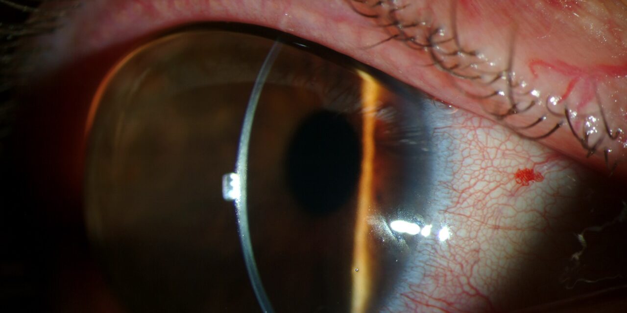
Megalocornea is a rare, non-progressive, bilateral condition in which the cornea is enlarged (generally defined as a horizontal diameter of 13 mm or greater) but otherwise structurally and functionally normal.
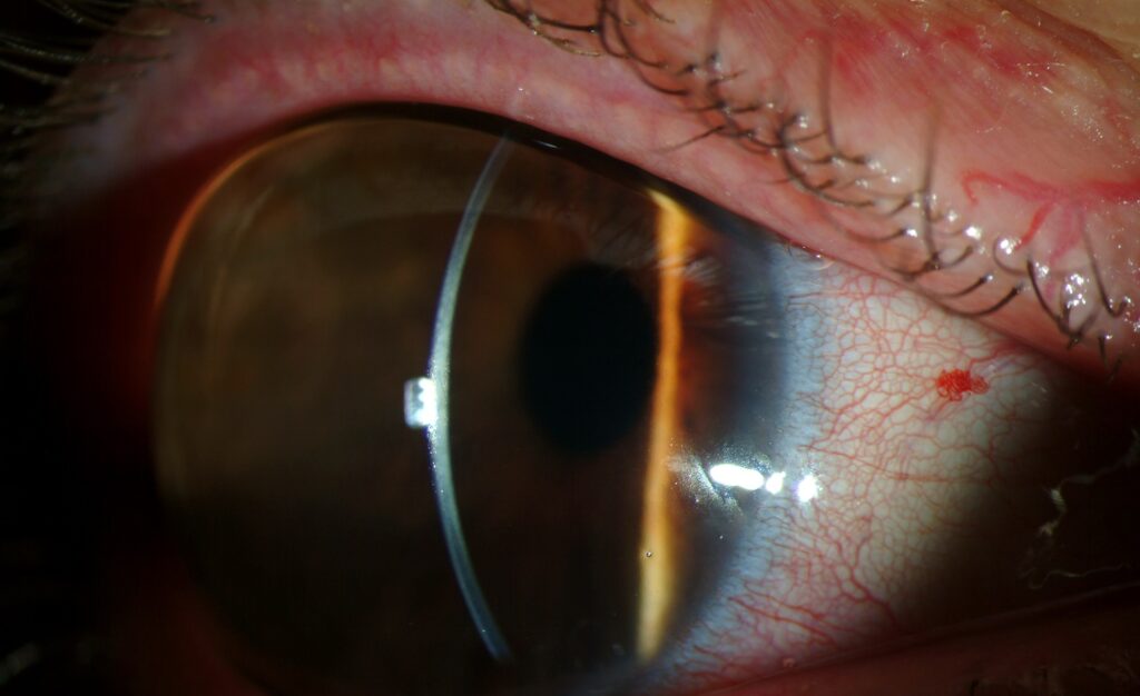
Its central clinical importance lies less in the corneal finding itself — which by definition causes no problems in isolation — rather than in the urgent need to distinguish it confidently from congenital glaucoma, which also enlarges the cornea but through an entirely different, actively damaging mechanism requiring emergency treatment rather than reassurance and observation.
Why “Normal but Large” Matters So Much
The defining feature of true megalocornea is that, apart from its size, the cornea is entirely normal — clear, of normal thickness, with a normal endothelium and no Haab’s striae — and intraocular pressure is normal, distinguishing it sharply from congenital glaucoma, in which corneal enlargement results from progressive stretching under chronically elevated pressure and is accompanied by the corneal clouding, breaks in Descemet’s membrane, and optic nerve damage that make that condition an ophthalmic emergency.
Confirming this distinction, rather than assuming either diagnosis based on corneal size alone, is the single most important diagnostic task in any infant or child presenting with large corneas.
Getting this wrong in either direction has real consequences: treating true megalocornea as glaucoma exposes a healthy eye to unnecessary intervention, while missing genuine congenital glaucoma delays sight-saving treatment during a period when timing critically affects the child’s long-term visual outcome.
Genetics and Associations
Most cases of isolated megalocornea are X-linked, predominantly affecting males, related to mutations in CHRDL1, though autosomal forms have also been described, and family history can be a useful clue when the inheritance pattern is apparent.
Megalocornea can occur as an isolated finding or as part of a broader systemic syndrome, including certain connective tissue disorders and Marfan syndrome, discussed in this site’s coverage of Marfan-related ectopia lentis, where megalocornea can be one of several ocular findings alongside the more classically emphasized lens subluxation, making a search for other suggestive systemic features worthwhile whenever the corneal finding is first identified.
All-fit Slit-Lamp Adapter
Record and share exactly what you see at the slit lamp. One adapter fits any slit lamp or surgical microscope — and any smartphone.
From Choroida — the team behind this siteAssociated Ocular Findings
- A deep anterior chamber, related to the overall enlarged anterior segment dimensions
- Iridodonesis (a fine tremulousness of the iris on eye movement), from relatively attenuated zonular support in an enlarged eye, sometimes accompanied by a degree of lens subluxation
- Increased risk of cataract, including presenile cataract developing earlier than typical age-related cataract, in a meaningful subset of patients
- Myopia, related to the altered ocular dimensions
- A mild increase in the risk of angle-closure glaucoma in adulthood in some patients, related to the altered anterior segment anatomy, despite the intraocular pressure being entirely normal at the time megalocornea itself is first identified in childhood
Differential Diagnosis
- Congenital glaucoma (buphthalmos) — elevated intraocular pressure, corneal clouding, Haab’s striae, and progressive rather than stable, non-progressive corneal enlargement, discussed in its own dedicated article on this site; this is the essential, urgent distinction to make in every case
- Keratoglobus — a diffusely thin, globular, ectatic cornea rather than an enlarged but structurally normal-thickness one, with its own distinct risk of spontaneous rupture from minor trauma, discussed in its own dedicated article on this site
- Anterior megalophthalmos — a broader term sometimes used when the entire anterior segment, not just the cornea, is proportionately enlarged, generally considered part of the same overall clinical spectrum as isolated megalocornea
Diagnostic Evaluation
Careful measurement of corneal diameter, intraocular pressure (often requiring examination under anesthesia in an infant, given how unreliable awake measurement can be at that age), corneal clarity, and corneal thickness (pachymetry) together establish that the enlarged cornea is stable and non-glaucomatous rather than progressively enlarging under elevated pressure.
Because a single exam cannot always fully exclude early or mild glaucoma with complete confidence, especially in a young infant, some clinicians favor a period of close follow-up with serial measurements before fully settling on a diagnosis of isolated, benign megalocornea, when any single finding (intraocular pressure, corneal clarity) is borderline rather than reassuring.
Given the association with Marfan syndrome and other connective tissue disorders, a general systemic and family history — tall stature, joint hypermobility, cardiac history — is a reasonable part of the initial evaluation.
A dilated exam specifically assessing for lens subluxation is worthwhile given the documented association between megalocornea and zonular laxity, and this same dilated exam is a reasonable opportunity to establish a baseline retinal and lens assessment for future comparison.
Management
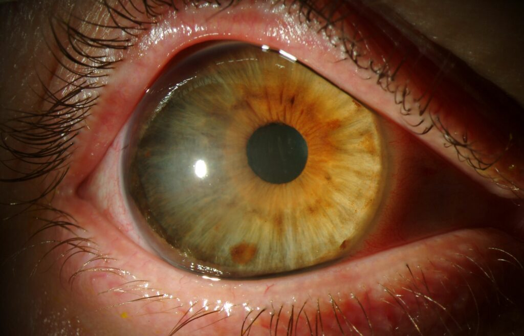
Isolated, uncomplicated megalocornea requires no specific treatment for the corneal finding itself.
Management centers on regular monitoring, given the modestly elevated long-term risks of cataract, lens subluxation, and angle-closure glaucoma described above, along with appropriate refractive correction for any associated myopia.
Any associated cataract or lens subluxation is managed according to the same general principles used for these conditions from other causes, discussed in this site’s separate coverage of both cataract and ectopia lentis, though surgical planning in an eye with megalocornea’s altered anatomy requires specific attention to the unusual anterior segment dimensions.
Surgeons operating on these eyes need to account for the larger-than-typical corneal diameter and deeper anterior chamber when selecting instrumentation and intraocular lens power, since standard assumptions calibrated to average-sized eyes may not directly transfer to this atypical anatomy.


Document what you see
Two smartphone imaging tools built for everyday clinic use — one for the slit lamp, one for the fundus.
From Choroida — the team behind this siteReferences
- Skuta GL, Sugar J, Ericson ES. Corneal endothelial cell measurements in megalocornea. Archives of Ophthalmology.
- Meire FM. Megalocornea. Clinical and genetic aspects. Documenta Ophthalmologica.
- American Academy of Ophthalmology. Basic and Clinical Science Course, Section 6: Pediatric Ophthalmology and Strabismus.
