
Medulloepithelioma is a rare intraocular tumor arising from the primitive, non-pigmented ciliary epithelium — embryologically the same tissue that gives rise to the neural retina — and it is overwhelmingly a disease of young children, with a typical age at diagnosis well below that of retinoblastoma, the far more common childhood intraocular tumor it can occasionally be mistaken for.
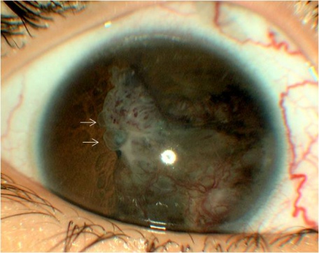
Because it arises from the ciliary body rather than the retina, its clinical presentation and imaging findings differ meaningfully from retinoblastoma despite both being pediatric intraocular malignancies, and correctly distinguishing the two matters directly for how the tumor is imaged, staged, and ultimately treated.
Origin and Classification
The tumor is classified as teratoid or non-teratoid based on whether it contains heteroplastic elements (such as cartilage) in addition to the neuroepithelial tissue that defines the tumor, and as benign or malignant based on histologic features including degree of cellular differentiation and invasive behavior.
Despite the word “malignant” applying to a subset of cases, even malignant medulloepithelioma has a low rate of distant metastasis compared with retinoblastoma, with local, intraocular tissue destruction and secondary complications being the more immediate clinical concern usually.
Clinical Presentation
- Decreased vision, often the presenting complaint, related to the tumor’s growth and its secondary effects on nearby structures
- Pain, frequently from secondary glaucoma as the growing ciliary body mass distorts the angle and impairs aqueous outflow
- A visible ciliary body or iris mass in some cases, sometimes associated with iris neovascularization
- Cataract, from direct tumor involvement of the lens or lens-related structures given the tumor’s origin adjacent to the ciliary body and zonules
- Leukocoria, in some cases, which is what most often prompts initial concern for retinoblastoma given the shared presenting sign in a young child, despite the two tumors arising from entirely different tissue
Fundus Explorer Pro
Photograph the retinal findings described here with the phone already in your pocket — 22 D optics and built-in illumination in one handheld unit.
From Choroida — the team behind this siteWhy It’s Often Diagnosed Late
Because medulloepithelioma arises from the ciliary body — a structure hidden behind the iris and not directly visible on routine external or even standard fundus examination — it is frequently not recognized until it has grown large enough to produce secondary findings (cataract, glaucoma, or a visible mass) rather than being caught at an earlier, more occult stage the way some other intraocular tumors might be.
This anatomic hiddenness is a genuine and recurring theme in the diagnostic delay commonly reported for this tumor.
Because the delay itself allows the tumor more time to grow and produce secondary complications before being recognized, a lower threshold for ultrasound biomicroscopy or examination under anesthesia in a young child with otherwise unexplained unilateral cataract, glaucoma, or a partial view of the fundus is genuinely worthwhile, rather than attributing these findings to a more common, benign cause by default.
Differential Diagnosis
- Retinoblastoma — the most important alternative diagnosis given overlapping presenting signs (leukocoria, glaucoma) in a similar young age group, discussed in its own dedicated article on this site; distinguished by the ciliary body (rather than retinal) origin and location on careful exam and imaging, along with some differences in typical presenting age and growth pattern
- Persistent fetal vasculature — also causes leukocoria and cataract in infancy, but with characteristic fibrovascular stalk findings and a different, non-neoplastic mechanism, discussed in its own dedicated article on this site
- Coats disease — leukocoria from exudative retinal changes rather than a discrete ciliary body mass, discussed in its own dedicated article on this site
- Iris or ciliary body cyst — generally translucent rather than solid on ultrasound biomicroscopy, without the more heterogeneous, solid appearance typical of medulloepithelioma
Diagnostic Evaluation
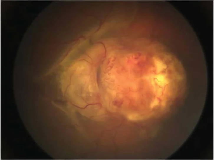
Ultrasound biomicroscopy is valuable for characterizing a ciliary body mass in detail, given the structure’s location behind the iris where direct visualization is limited, and can reveal cystic spaces within the tumor that are a characteristic feature of medulloepithelioma.
MRI helps assess the full extent of the tumor and any extraocular extension, an important consideration for both prognosis and surgical planning.
Examination under anesthesia is often required in a young child to allow the detailed anterior segment and ciliary body assessment needed, given how difficult a fully cooperative exam is at this age, and imaging is frequently obtained during the same session to minimize the number of separate anesthetic exposures.
Management
Enucleation remains the standard treatment for most cases, larger tumors or those with malignant features, given the tumor’s location and the technical difficulty of achieving complete, margin-negative local resection in the ciliary body region without unacceptable risk of incomplete removal.
Local resection (iridocyclectomy) is considered for select, smaller, more anteriorly located tumors in centers with appropriate ocular oncology surgical expertise, aiming to preserve the eye when the tumor’s size and location make this a reasonable option without compromising oncologic safety.
Because distant metastasis, while less common than in retinoblastoma, does occur in malignant medulloepithelioma, appropriate staging and systemic follow-up is part of comprehensive management once the diagnosis is confirmed, generally coordinated with pediatric oncology given the rarity and complexity of the tumor.
Given how uncommon this tumor is, management is best concentrated at centers with specific ocular oncology expertise, where the pathologist, surgeon, and oncology team have enough collective experience with this rare entity to guide decisions that a more general practice setting might not routinely encounter.


Document what you see
Two smartphone imaging tools built for everyday clinic use — one for the slit lamp, one for the fundus.
From Choroida — the team behind this siteReferences
- Broughton WL, Zimmerman LE. A clinicopathologic study of 56 cases of intraocular medulloepitheliomas. American Journal of Ophthalmology.
- Kaliki S, Shields CL, Eagle RC Jr, et al. Ciliary body medulloepithelioma: analysis of 41 cases. Ophthalmology.
- American Academy of Ophthalmology. Basic and Clinical Science Course, Section 6: Pediatric Ophthalmology and Strabismus.
Test yourself
A few questions straight from this article.
-
From which tissue does intraocular medulloepithelioma arise?
The tumour arises from primitive non-pigmented ciliary epithelium, embryologically the same tissue that gives rise to the neural retina. -
What makes a medulloepithelioma teratoid rather than non-teratoid?
Teratoid tumours contain heteroplastic elements, cartilage among them, alongside the neuroepithelial tissue that defines the tumour. -
Why is pain a common presenting symptom of ciliary body medulloepithelioma?
The growing ciliary body mass distorts the angle and impairs aqueous outflow, and the resulting secondary glaucoma frequently causes pain. -
Why is medulloepithelioma often diagnosed late?
The ciliary body is not directly visible on routine external or standard fundus examination, so the tumour is often recognised only once it causes cataract, glaucoma or a visible mass. -
A young child has unexplained unilateral cataract and glaucoma. Which approach does this warrant?
Attributing unexplained unilateral cataract, glaucoma or a partial fundus view to a commoner benign cause risks further delay, so imaging the ciliary body early is worthwhile. -
Which ultrasound biomicroscopy feature is characteristic of medulloepithelioma?
Ultrasound biomicroscopy can reveal cystic spaces within the lesion, a characteristic feature of medulloepithelioma, and characterises a mass hidden behind the iris. -
Which appearance on ultrasound biomicroscopy suggests a ciliary body cyst rather than medulloepithelioma?
Iris and ciliary body cysts appear translucent, lacking the more heterogeneous, solid appearance typical of medulloepithelioma. -
Why does enucleation remain the standard treatment for most medulloepitheliomas?
Achieving complete, margin-negative local resection in the ciliary body region is technically difficult without unacceptable risk of incomplete removal, particularly in larger tumours or those with malignant features. -
Which tumours may be considered for local resection by iridocyclectomy instead of enucleation?
Eye-preserving iridocyclectomy is considered for select smaller, anteriorly located tumours in centres with ocular oncology surgical expertise, provided oncologic safety is not compromised. -
How does the metastatic behaviour of malignant medulloepithelioma compare with retinoblastoma?
Local intraocular tissue destruction is the more immediate concern, but because distant metastasis does occur, staging and systemic follow-up with paediatric oncology remain part of management.
