
Neurotrophic keratopathy is a degenerative corneal disease that develops when the trigeminal nerve’s sensory innervation of the cornea is impaired, removing not just pain sensation but also the trophic support the nerve normally provides to corneal epithelial cells.
The result is a cornea that heals poorly, breaks down easily, and — because the patient cannot feel the damage happening — frequently progresses further before anyone notices than it would if pain were still driving the patient to seek care.
Why Corneal Nerves Matter Beyond Sensation
Corneal sensory nerves release neuropeptides (including substance P and calcitonin gene-related peptide) that support normal epithelial cell proliferation, migration, and adhesion, independent of their role in transmitting pain and touch.
When this trophic input is lost, epithelial healing slows, and epithelial integrity becomes fragile even without any additional insult.
The cornea essentially loses part of its normal maintenance and repair machinery, not just its alarm system.
Causes
- Herpes simplex and herpes zoster keratitis — among the most common causes, from direct viral damage to corneal nerve fibers
- Prior intracranial or skull base surgery involving the trigeminal nerve, including surgery for trigeminal neuralgia or acoustic neuroma
- Diabetes mellitus, through diabetic corneal neuropathy, often underrecognized as a contributing factor
- Chemical burns and other injuries that damage corneal nerve endings directly
- Contact lens overwear
- Topical medication toxicity from chronic use of certain preserved drops
- Congenital causes, including congenital corneal anesthesia and certain rare genetic syndromes affecting trigeminal nerve development
- Leprosy, where trigeminal nerve involvement is a recognized contributor to corneal disease, discussed in more detail in the dedicated article on leprosy’s ocular findings on this site
All-fit Slit-Lamp Adapter
Record and share exactly what you see at the slit lamp. One adapter fits any slit lamp or surgical microscope — and any smartphone.
From Choroida — the team behind this siteClinical Staging
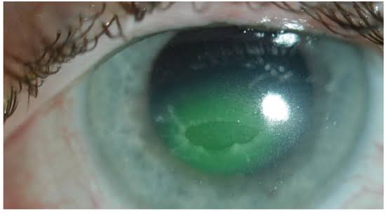
Neurotrophic keratopathy is classically staged from mild (stage 1: epithelial changes — irregularity, punctate keratopathy, reduced tear film stability — without a frank defect) through moderate (stage 2: a persistent epithelial defect, often with rolled, thickened edges that fail to progress toward healing despite standard measures) to severe (stage 3: stromal involvement with thinning, melting, and a genuine risk of descemetocele formation or perforation).
This staging directly informs the intensity of treatment, because stage 1 disease can often be managed with lubrication alone while stage 3 disease requires the same urgency as any other cause of impending corneal perforation.
Clinical Findings
- Reduced or absent corneal sensation on formal testing (cotton wisp or, more precisely, Cochet-Bonnet esthesiometry) — the single most important and specific finding, and one that should be actively checked whenever the diagnosis is suspected
- A persistent epithelial defect, often oval or round, with characteristically rolled, gray, thickened edges rather than the clean margins of a simple abrasion
- Notably little pain or discomfort relative to the severity of the corneal finding — a mismatch that is itself diagnostically important
- Reduced or absent blink reflex on the affected side in some cases
- Stromal thinning or melting in advanced disease
Differential Diagnosis
- Ordinary persistent epithelial defect from dry eye or exposure — corneal sensation is typically preserved, distinguishing it from a neurotrophic cause
- Infectious keratitis — usually more painful, with an infiltrate rather than isolated epithelial breakdown, though secondary infection of a neurotrophic defect is itself a real risk requiring ongoing vigilance
- Recurrent corneal erosion — episodic, typically with normal corneal sensation and a history of prior abrasion or an underlying epithelial basement membrane dystrophy
Management
Preservative-free lubrication is the foundation of treatment at every stage, both to protect the fragile ocular surface and to reduce the toxic load from preservatives that a normally functioning cornea would tolerate better.
Autologous serum or plasma eye drops, which supply growth factors and neurotrophic support the cornea would otherwise get from intact innervation, have become an important treatment for persistent defects.
Recombinant human nerve growth factor (cenegermin) is now available specifically for neurotrophic keratopathy and has meaningfully changed outcomes for many patients with moderate to severe disease.
Bandage contact lenses, amniotic membrane transplantation, and temporary or permanent tarsorrhaphy all serve to protect the ocular surface and support healing in stage 2 and 3 disease, with the choice depending on defect severity and response to less invasive measures.
Any secondary microbial infection needs to be treated aggressively given the impaired healing capacity of the underlying cornea.
Eyes progressing to descemetocele or perforation are managed with the same urgency described for those findings elsewhere on this site.


Document what you see
Two smartphone imaging tools built for everyday clinic use — one for the slit lamp, one for the fundus.
From Choroida — the team behind this siteReferences
- Bonini S, Rama P, Olzi D, Lambiase A. Neurotrophic keratitis. Eye.
- Sacchetti M, Lambiase A. Diagnosis and management of neurotrophic keratitis. Clinical Ophthalmology.
- Bonini S, Lambiase A, Rama P, et al. Phase II randomized, double-masked, vehicle-controlled trial of recombinant human nerve growth factor for neurotrophic keratitis. Ophthalmology.
- American Academy of Ophthalmology. Basic and Clinical Science Course, Section 8: External Disease and Cornea.
