
Age-related macular degeneration (AMD) is a progressive eye condition that affects the macula, a small but crucial part of the retina responsible for central vision.
As AMD advances, it can lead to severe vision impairment and impact the quality of life for affected individuals. Managing AMD involves a combination of lifestyle changes, medical interventions, and supportive therapies.
In this comprehensive guide, we’ll explore various AMD management strategies.
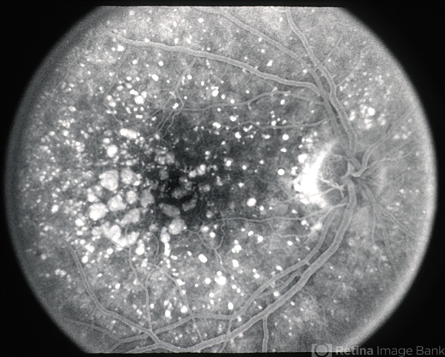
Age-Related Macular Degeneration Medical therapy
Non-neovascular ARMD Treatments
Antioxidant and mineral supplementation:
Antioxidant and mineral supplementation has been shown to reduce the risk of progression in ARMD. The daily amounts of antioxidants and zinc in the AREDS formulation are:
- 500 milligrams of vitamin C
- 400 International Units of vitamin E
- 15 milligrams of beta-carotene (equivalent to 25,000 International Units of vitamin A)
- 80 milligrams of zinc as zinc oxide
- 2 milligrams of copper as cupric oxide
Since high-dose zinc supplementation can lead to copper-deficiency anemia by inhibiting copper absorption at the level of the enterocyte, copper supplementation was added to the AREDS formula.
Cigarette smokers should be warned about the small, but real potential risk of lung cancer with high-dose beta-carotene supplementation in the AREDS formula.
Non-beta-carotene-containing supplements are more appropriate for this subgroup of patients.
Based on data suggesting that increasing the intake of lutein + zeaxanthin, and omega-3 long-chain polyunsaturated fatty acids (docosahexaenoic acid [DHA] + eicosapentaenoic acid [EPA]) might reduce the risk of developing advanced AMD, AREDS2, a randomized, double-masked control trial was started.
AREDS2 was designed to test whether adding lutein + zeaxanthin, DHA + EPA, or lutein + zeaxanthin + DHA + EPA to the AREDS formulation further reduces the risk of progression to advanced AMD.
Another goal of AREDS was to test the effects of eliminating beta carotene and reducing the zinc dose from the AREDS formulation. The following are the modifications of AREDS2:
- 10 mg lutein and 2 mg zeaxanthin
- 350 mg DHA and 650 mg EPA
- No beta-carotene
- 25 mg zinc
In AREDS2, lutein/zeaxanthin or DHA/EPA had no additional effect on the risk of advanced AMD. Study participants who took AREDS containing lutein/zeaxanthin and no beta-carotene had a slight reduction in the risk of advanced AMD, compared to those who took AREDS with beta-carotene and no lutein/zeaxanthin.
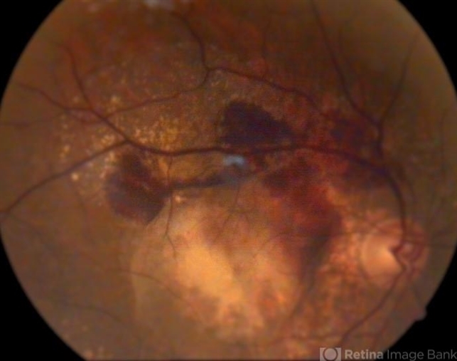
Importantly, former smokers who took AREDS with beta-carotene had a higher incidence of lung cancer. Lower zinc oxide doses (25mg) did not significantly increase the risk of advanced AMD, although a trend to more protection from advanced AMD was noted with higher zinc oxide doses (80mg).
Complement Factor Inhibitors:
The complement factor 3 inhibitor pegcetacoplan has been assessed for the treatment of geographic atrophy in the phase 3 DERBY and OAKS trials.
In the DERBY trial, monthly dosing of pegcetacoplan led to a 36% decrease in lesion growth, while every other month dosing of pegcetacolplan led to a 29% decrease in lesion growth at 24 months.
The OAKS trial showed a 24% decrease in GA growth with every month dosing, and a 25% decrease in lesion growth with every other month dosing. No clinically meaningful differences were noted in functional outcomes.
In terms of adverse events in DERBY and OAKS, the monthly pegcetacoplan had a 12% rate of choroidal neovascularization (CNV), 3.8% risk of intraocular inflammation (IOI), and 1.7% rate of ischemic optic neuropathy, while the every other month dose had a 7% rate of CNV, 2.1% rate of IOI, and 0.2% of optic neuropathy, compared to a 3% risk of CNV, 0.2% risk of IOI, and 0% risk of optic neuropathy in the sham.
The complement factor 5 inhibitor avacincaptad pegol has also been assessed for the treatment of geographic atrophy in the phase 2/3 GATHER1 and phase 3 GATHER2 trials.
GATHER1 evaluated multiple doses of avacincaptad, the first part looked at 1 and 2 mg monthly versus sham in a 1:1:1 ratio, and the second part compared 2 and 4 mg monthly versus sham in a 1:1:1 ratio.
GATHER2 evaluated 2 mg avacincaptad monthly versus sham in a 1:1 ratio, again with primary endpoint of mean rate of growth of GA at 12 months and with secondary endpoints at 18 months related to GA growth and visual function.
The inclusion criteria are notable for requiring non-subfoveal GA, thus these patients did not have subfoveal GA in contrast to the OAKS and DERBY trials. At 12 months, GATHER1 showed a 35% decrease in GA growth, while GATHER 2 showed a 17.7% decrease in GA growth.
No statistically significant effects were found in BCVA between treated and SHAM groups. The 2mg dose of avacincaptad in GATHER1 had a 9% risk of CNV, 1.5% of IOI, and no instance of optic neuropathy compared to a 2.7% risk of CNV in the sham.
In GATHER2, there was a 6.7% risk of CNV vs a 4.1% risk in sham, and no instances of IOI or optic neuropathy. Avacincaptad pegol was approved for the treatment of GA by the FDA on August 6, 2023.
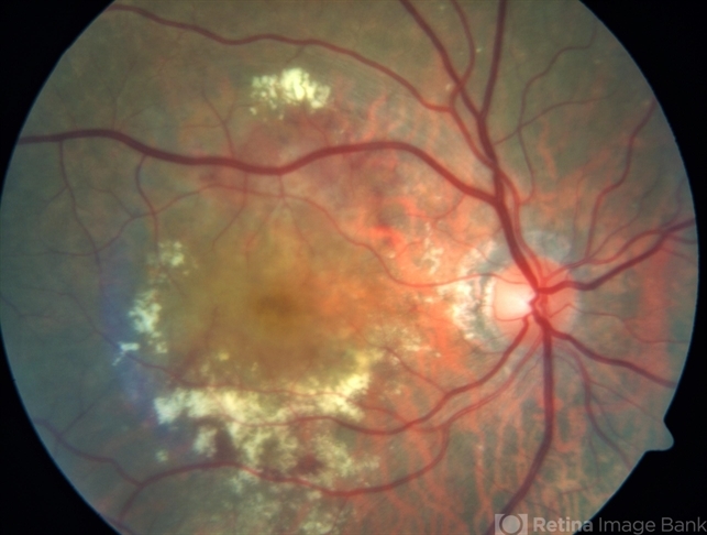
Neovascular ARMD Treatments
Macular Photocoagulation Studies:
Studies performed in the 1980s assessed the efficacy of laser photocoagulation in limiting the damage caused by choroidal neovascular lesions. These studies evaluated laser treatment of extrafoveal, juxtafoveal, and subfoveal neovascular membranes.
Patients with direct laser photocoagulation to extrafoveal or juxtafoveal sites fared better than those receiving direct laser to subfoveal membranes.
While severe vision loss was averted, laser photocoagulation currently has limited utility because of its high recurrence rates, risk of inducing vision loss (especially with subfoveal membranes), and failure to affect an improvement in vision from baseline visual acuity.
In the era of anti-VEGF therapies, the indications for direct photocoagulation are diminishing.
However, it may be considered in extrafoveal small CNVM.
verteporfin Photodynamic therapy/vPDT:
The use of photosensitizers (e.g., verteporfin) that pool in neovascular membranes and subsequently produce reactive oxygen species upon activation with light of a specific wavelength was introduced in the late 1990s.
In the era of anti-VEGF therapies, the indications for photodynamic therapy are diminishing.
However, PDT has a definite role in the management of polypoidal choroidal vasculopathy and central serous chorioretinopathy.
EVEREST trial showed that ‘after 12 months, combination therapy of ranibizumab plus vPDT was not only non-inferior but also superior to ranibizumab monotherapy in best-corrected visual acuity and superior in complete polyp regression while requiring fewer injections’
Anti-VEGF treatments:
Optical coherence tomography and anti-vascular endothelial growth factor (VEGF) therapy together have revolutionized the treatment of exudative ARMD.
Ranibizumab (Lucentis, Genentech, San Francisco, CA), bevacizumab (Avastin, Genentech, San Francisco, CA), and aflibercept (Eylea, Regeneron Pharmaceuticals Inc., Tarrytown, NY), are frequently used in the treatment of exudative ARMD. Pegaptanib (Macugen, Pfizer) was the first anti-VEGF therapy to receive FDA approval for the treatment of ARMD in 2004.
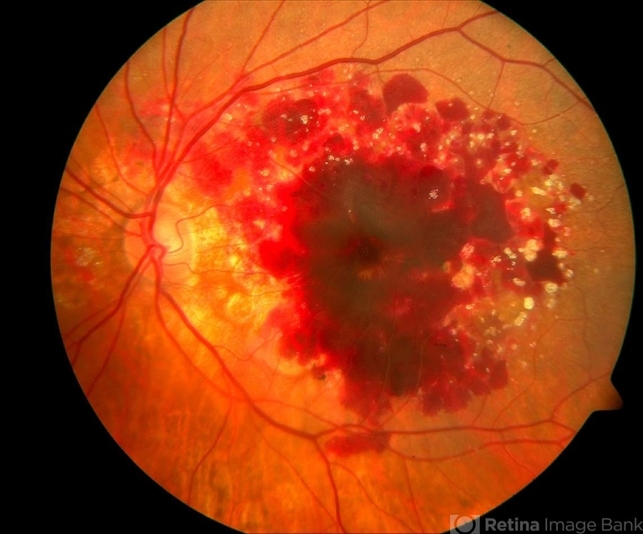
It is biochemically distinct from subsequent anti-VEGF agents, in that it represents an aptamer as opposed to a monoclonal antibody (bevacizumab), monoclonal antibody fragment (ranabizumab), or a receptor-antibody fusion protein (aflibercept, see diagram below).
Pegaptanib is a small oligonucleic acid molecule that binds specifically to the VEGF-165 isoform.
The most recent anti-VEGF agent that was recently approved was brolucizumab (Beovu, Novartis, Cambridge, MA), which is a single-chain antibody fragment against VEGF, which was found to be non-inferior to aflibercept, and more than half of patients were able to extend to 3 months dosing with brolucizumab within the first year as demonstrated in the HAWK and HARRIER trials.
In an effort to extend dosing, a new port delivery system was developed by Genentech to deliver continuous levels of ranibizumab.
The ranibizumab port delivery system (Susvimo, Genentech, San Francisco, CA) is a unique surgical implant that is placed within the vitreous and provides continuous delivery of ranibizumab.
This implant was approved by the FDA in October 2021, and this delivery system can be refilled every 6 months.
The Archway study showed the non-inferiority of the port delivery system compared to monthly ranibizumab injections. 98% of patients were able to go 6 months before their first refill.
Age-Related Macular Degeneration Radiation therapy
Radiotherapy has been used in neovascular ARMD under the premise that radiation can inhibit the exuberant cellular proliferation necessary to create the choroidal neovascular membrane.
However, studies on efficacy have not shown a clear benefit of this treatment modality.
Anti-VEGF and Anti-Ang2 treatment:
The first bi-specific antibody therapy for the treatment of neovascular ARMD was recently approved by the FDA on January 31, 2022.
Faricimab (Vabysmo, Genentech, San Francisco, CA) is a bi-specific antibody directed against both VEGF and angiopoietin-2.
This dual-targeted treatment was dosed up to 4-month intervals and was found to be non-inferior to aflibercept given every 2 months in the first year.
In the TENAYA and LUCERNE trials, a large majority of patients could be dosed at 3-month intervals or more, showing improved efficacy and durability of the treatment compared to the anti-VEGF inhibition alone.
Fundus Explorer Pro
Photograph the retinal findings described here with the phone already in your pocket — 22 D optics and built-in illumination in one handheld unit.
From Choroida — the team behind this siteAge-Related Macular Degeneration Surgery
Submacular surgery in ARMD has included approaches to translocate the macula, either by vitrectomy and retinotomy or by vitrectomy and sclerochroidal foreshortening (achieved externally), yet these procedures have not gained widespread use.
In instances where a large submacular hemorrhage occurs, pneumatic displacement with face-down positioning and intraocular gas injection can be performed.
Some have used vitrectomy, retinotomy, and subretinal injection of tissue plasminogen activator (tPA) in instances of large submacular hemorrhage.
The NEI-funded Submacular Surgery Trials (SST) evaluated the visual acuity outcome and complications of excising choroidal neovascular membranes.
In patients with large central macular, subretinal hemorrhage due to choroidal neovascularization (new or recurring after laser photocoagulation; Group B protocol), there were no significant differences in BCVA between the surgery and observation arms of the study.
The surgical intervention can vary from patient to patient, but it has the goal of removing the entire neovascular membrane, any blood, and any scar tissue present.


Document what you see
Two smartphone imaging tools built for everyday clinic use — one for the slit lamp, one for the fundus.
From Choroida — the team behind this siteReferences
- Bressler SB, Do DV, Bressler NM. Age-related macular degeneration: drusen and geographic atrophy. In: Albert DM, Miller JW, Azar DT, Blodi BA, eds. Albert and Jakobiec’s Principles and Practice of Ophthalmology. 3rd ed. Philadelphia: Saunders; 2008: ch. 144.
- Lim LS, Mitchell P, Seddon JM, Holz FG, Wong TY (2012). Age-related macular degeneration. The Lancet 379:1728-1738.
- Huang D, Swanson EA, Lin CP, Shuman JS, Stinson WG, Chang W, Hee MR, Flotte T, Gregory K, Puliafito CA, et al. 1991. Optical coherence tomography. Science 254 (5035): 1178-1181.
- Yannuzzi LA, Wong DW, Sforzolini BS, et al. Polypoidal choroidal vasculopathy and neovascularized age-related macular degeneration. ARch Ophthalmol. 1999; 117: 1503-1510.
- Bressler NM, Bressler SB, Sarks SH, Sarks JP. Chapter 60. Age-Related Macular Degeneration: Nonneovascular Early AMD, Intermediate AMD, and Geographic Atrophy, Volume II, 4th Edition. Elsevier, Mosby. 2006. Editor: Andrew SP. Schachat.
- Scripsema, N.K., Hu, D.N. and Rosen, R.B., 2015. Lutein, Zeaxanthin, and meso-Zeaxanthin in the Clinical Management of Eye Disease. Journal of Ophthalmology, 2015.
