
DISEASE
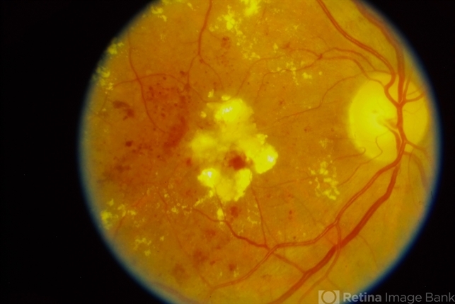
Diabetic retinopathy represents microvascular end-organ damage as a result of diabetes. It ranges from non-proliferative diabetic retinopathy (NPDR) and its stages to proliferative diabetic retinopathy (PDR).
As the disease progresses, associated diabetic macular edema (DME) may also become apparent. Among patients aged 25-74, diabetic retinopathy is a leading cause of vision loss worldwide.
By 2030 an estimated 191.0 million people globally will have diabetic retinopathy, and approximately 56.3 million will have vision-threatening diabetic retinopathy.
The Wisconsin Epidemiologic Study of Diabetic Retinopathy (WESDR) Cohort showed that after 20 years of diabetes mellitus, 99% of patients with type 1 and 60% of patients with type 2 show some degree of retinopathy.
There are several other key risk factors for the development of diabetic retinopathy beyond years since diagnosis and type of diabetes.
Additionally, elevated hemoglobin A1c (HbA1c) levels and blood pressure are associated with increased risk of diabetic retinopathy.
Type 1 diabetes mellitus (T1DM) is characterized by the destruction of beta cells in the pancreas by an autoimmune mechanism, whereas type 2 diabetes mellitus (T2DM) is a relationship between lifestyle and genetics.
The etiology of these two subtypes describes the etiology of diabetic retinopathy, as the retinal disease is an end-organ manifestation of the principal disease.
There is a stronger genetic association between T2DM compared to T1DM. Multiple genetic factors have been named in the development of T2DM including TCF7L2, NOTCH2, KCNQ1, JAZF1, and MODY (a heterogeneous disorder with autosomal dominant transmission). Poor lifestyle, in conjunction with genetic influences, increases the risk of developing T2DM.
Over 90% of people with a new diagnosis of T1DM have measurable antibodies against specific pancreatic β-cell proteins (insulin, islet antigen 2, zinc transporter 8, etc.).
These antibodies lead to a chain of progressive loss of β-cells, decreased insulin release, and recognizable diabetes.
The main types of diabetic retinopathy are non-proliferative diabetic retinopathy (NPDR) and proliferative diabetic retinopathy (PDR).
The distinguishing feature between these two categories is the presence (proliferative) or absence (non-proliferative) of abnormal new blood vessels (retinal, optic disc, or iris/angle neovascularization).
Of primary concern are the factors that lead to visual impairment in this patient population. The three items listed below are the foundation of this disease process, and their presence can be correlated with disease severity.
- Capillary leakage (DME)
- Capillary occlusion
- Sequelae of retinal ischemia (retinal neovascularization, vitreous hemorrhage, tractional retinal detachment, neovascular glaucoma)
Diagnosis
History:
Symptoms of decreased vision or fluctuating vision (lens or macular edema), presence of floaters (vitreous hemorrhage), or visual field defects (tractional detachment). It is important to know the hemoglobin A1c and whether or not the patient’s blood pressure is under control.
Physical Examination and Signs:
A slit lamp examination and dilated fundus examination should be performed. One should look carefully for the presence of abnormal blood vessels on the iris [neovascularization of the iris (NVI) or rubeosis], cataracts (associated with diabetes), and vitreous cells (blood in the vitreous or pigmented cells if there is a retinal detachment with hole formation).
Intraocular pressure (IOP) should be checked especially when NVI is seen. Dilated fundus examination should include a macular examination (contact lens or non-contact lens) to look for microaneurysms, hemorrhage, hard exudates, cotton wool spots, and retinal swelling (DME).
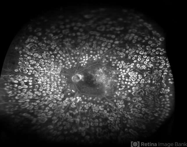
The optic disc and area surrounding it (for one disc diameter) should be examined for the presence of abnormal new blood vessels (neovascularization of the disc, NVD), optic nerve head pallor, or glaucomatous changes.
The remainder of the retina should also be examined for the presence of abnormal new blood vessels (neovascularization elsewhere, NVE).
Fundus Explorer Pro
Photograph the retinal findings described here with the phone already in your pocket — 22 D optics and built-in illumination in one handheld unit.
From Choroida — the team behind this siteSymptoms
Clinical Diagnosis:
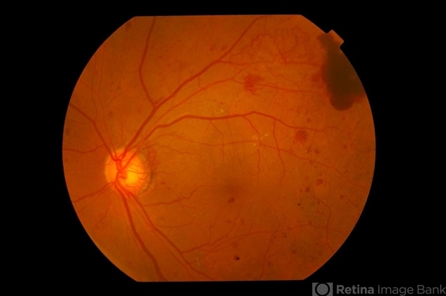
The central retinal area that is located between the main branches (superior and inferior arcades) of the central retinal vessels (central retinal artery and central retinal vein) in the eye is known as the “macular area.”
The retina beyond this is considered the “peripheral retina.” The central retinal area can develop abnormal findings in diabetic retinopathy.
These findings can be present in the non-proliferative or the proliferative forms of the disease. These changes in the macula include the presence of abnormally dilated small vessel outpouchings (called microaneurysms), retinal bleeding (retinal hemorrhages), and yellow lipid and protein deposits (hard exudates).
The macula can get thicker than normal, which is referred to as macular edema.

MANAGEMENT
General Treatment:
The systemic status should be optimized by
-
Strict metabolic control of diabetes,
-
Maintaining HbA1C levels under 7%,
-
Lifestyle modifications like routine exercises and a proper diabetic food diet,
Patients should visit diabetologists for proper follow-up visits and take timely antidiabetic medications.
Read more about management strategies
Other systemic ailments like hypertension, dyslipidemia, hypoproteinemia, anemia, nephropathy, neuropathy, cardiac ailments, and others should also be taken care of by respective medications and excellent interprofessional collaboration.


Document what you see
Two smartphone imaging tools built for everyday clinic use — one for the slit lamp, one for the fundus.
From Choroida — the team behind this siteREFERENCES
- Brown DM1, Nguyen QD, Marcus DM, Boyer DS, Patel S, Feiner L, Schlottmann PG, Rundle AC, Zhang J, Rubio RG, Adamis AP, Ehrlich JS, Hopkins JJ; RIDE and RISE Research Group.Long-term outcomes of ranibizumab therapy for diabetic macular edema: the 36-month results from two phase III trials: RISE and RIDE.Ophthalmology. 2013 Oct;120(10):2013-22. doi: 10.1016/j.ophtha.2013.02.034. Epub 2013 May 22.
- Davis MD, Fisher MR, Gangnon RE, Barton F, Aiello LM, Chew EY, Ferris FL 3rd, Knatterud GL. Risk factors for high-risk proliferative diabetic retinopathy and severe visual loss: Early Treatment Diabetic Retinopathy Study Report #18. Invest Ophthalmol Vis Sci. 1998 Feb; 39:233-52
- Diabetes Control and Complications Trial Research Group. Clustering of long-term complications in families with diabetes in the diabetes control and complications trial. Diabetes. 1997 Nov; 46:1829-39.
- Diabetic Retinopathy Clinical Research Network. A randomized trial comparing intravitreal triamcinolone acetonide and focal/grid photocoagulation for diabetic macular edema. Ophthalmology. 2008 Sep;115(9):1447-9, 1449.e1-10. Epub 2008 Jul 26.
- Diabetic Retinopathy Clinical Research Network (DRCR.net), Beck RW, Edwards AR, Aiello LP, Bressler NM, Ferris F, Glassman AR, Hartnett E, Ip MS, Kim JE, Kollman C. Three-year follow-up of a randomized trial comparing focal/grid photocoagulation and intravitreal triamcinolone for diabetic macular edema. Arch Ophthalmol. 2009 Mar;127(3):245-51.
- Diabetic Retinopathy Clinical Research Network, Elman MJ, Aiello LP, Beck RW, Bressler NM, Bressler SB, Edwards AR, Ferris FL 3rd, Friedman SM, Glassman AR, Miller KM, Scott IU, Stockdale CR, Sun JK. Randomized trial evaluating ranibizumab plus prompt or deferred laser or triamcinolone plus prompt laser for diabetic macular edema. Ophthalmology. 2010 Jun;117(6):1064-1077.e35. Epub 2010 Apr 28.
Test yourself
A few questions straight from this article.
-
Diabetic retinopathy represents which kind of damage caused by diabetes?
Diabetic retinopathy is microvascular end-organ damage resulting from diabetes, ranging through the stages of non-proliferative disease to proliferative diabetic retinopathy. -
Which single feature distinguishes proliferative from non-proliferative diabetic retinopathy?
The distinguishing feature is the presence or absence of abnormal new vessels, whether retinal, on the optic disc, or on the iris and in the angle. -
In the WESDR cohort, what proportion of type 1 diabetics showed retinopathy after 20 years of diabetes?
The Wisconsin Epidemiologic Study of Diabetic Retinopathy found that after 20 years of diabetes, 99% of type 1 patients and 60% of type 2 patients showed some degree of retinopathy. -
Which three processes form the foundation of visual impairment in diabetic retinopathy?
The three foundations are capillary leakage producing macular oedema, capillary occlusion, and the sequelae of retinal ischaemia such as neovascularisation, vitreous haemorrhage, tractional detachment and neovascular glaucoma. -
Type 1 diabetes mellitus, the substrate for diabetic retinopathy in younger patients, arises by which mechanism?
Type 1 diabetes is characterised by autoimmune destruction of pancreatic beta cells; over 90% of newly diagnosed patients carry measurable antibodies against beta-cell proteins such as insulin, islet antigen 2 and zinc transporter 8. -
Neovascularisation of the iris is found in a diabetic eye. Which measurement becomes especially important?
Intraocular pressure should be checked especially when neovascularisation of the iris, or rubeosis, is seen, since neovascular glaucoma is among the sequelae of retinal ischaemia. -
When examining for neovascularisation of the disc in diabetic retinopathy, how wide an area around the disc is assessed?
The optic disc and the area within one disc diameter of it are examined for new vessels on the disc, as well as for optic nerve head pallor and glaucomatous change. -
Which landmark bounds the macular area as defined for diabetic retinopathy assessment?
The macular area is the central retina lying between the superior and inferior arcades of the central retinal vessels; retina beyond this is the peripheral retina. -
A diabetic patient reports new floaters. Which complication does this symptom point toward?
Floaters point to vitreous haemorrhage, while visual field defects suggest tractional detachment and fluctuating vision suggests lens change or macular oedema. -
What HbA1c level is targeted in the general treatment of a patient with diabetic retinopathy?
Systemic status is optimised with strict metabolic control and HbA1c maintained under 7%, alongside exercise, a proper diabetic diet and regular diabetologist follow-up.

Writes and reviews the clinical articles on Educate. Every post is checked against peer-reviewed sources before it goes live.


