
Dengue maculopathy is a macular complication of dengue fever that causes central blurring or a scotoma in the days or weeks after the acute febrile illness.
Ocular involvement is a recognized but uncommon complication, and it usually appears around the time that platelet counts are low.
Most patients recover good vision, though some are left with residual scotomas from macular ischemia.
Clinicians in endemic regions, and those who see travelers returning from them, should keep the diagnosis in mind when a recent febrile illness is followed by central visual loss.
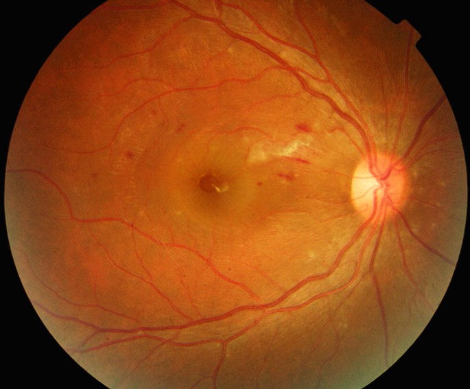
What Is Dengue Maculopathy?
Dengue is a mosquito-borne flavivirus infection transmitted by Aedes species.
It produces a spectrum from mild febrile illness to severe dengue with plasma leakage and hemorrhage.
The disease is endemic in tropical and subtropical regions, and it has spread to new areas as vectors expand, including parts of the Arabian Peninsula and East Africa.
Ocular involvement has been documented in a minority of hospitalized patients.
In a prospective study, dengue maculopathy was found in about 10 percent of patients admitted with dengue fever, though the frequency varies between studies and with the intensity of screening.
Pathogenesis
Several mechanisms may contribute.
- Direct viral effects on the retinal vascular endothelium
- Immune-mediated vasculitis and immune complex deposition
- Increased vascular permeability similar to that seen in systemic dengue
- Thrombocytopenia and coagulopathy, which contribute to hemorrhage
- Ischemia of the deep capillary plexus and the perifoveal capillary network
Ocular symptoms typically begin around the time of the nadir of the platelet count, and they are associated with lower platelet levels in some series, although they can occur in patients without severe thrombocytopenia.
Fundus Explorer Pro
Photograph the retinal findings described here with the phone already in your pocket — 22 D optics and built-in illumination in one handheld unit.
From Choroida — the team behind this siteClinical Presentation
Patients report blurred vision, a central or paracentral scotoma, metamorphopsia, or floaters.
The onset is usually within one to two weeks of the fever, and both eyes may be involved.
Fundus Findings
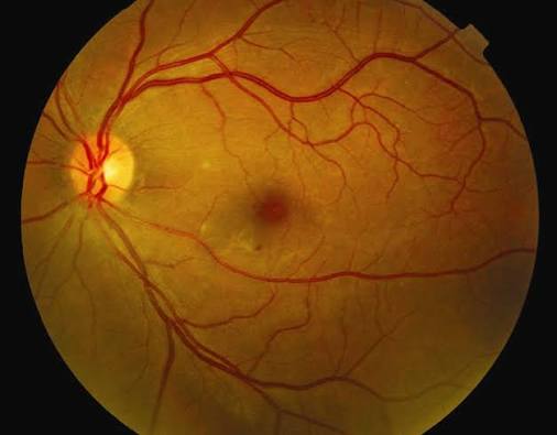
- Foveolitis, seen as a yellowish-white spot at the fovea
- Macular edema, and sometimes serous detachment
- Intraretinal and preretinal hemorrhages, including subhyaloid blood
- Cotton wool spots
- Retinal vasculitis with sheathing
- Retinal vascular occlusion
- Optic disc swelling or optic neuritis in some cases
Anterior uveitis, vitritis, and subconjunctival hemorrhage may accompany the posterior findings.
Imaging
OCT
OCT is the most helpful test.
Patterns include:
- Diffuse retinal thickening with cystoid spaces
- Hyperreflectivity and thickening of the outer retinal layers at the fovea, in the case of foveolitis
- Subretinal fluid
- Disruption of the ellipsoid zone
- Middle-layer hyperreflective bands, in keeping with deep capillary ischemia (see paracentral acute middle maculopathy)
In a study of dengue maculopathy, several OCT patterns were used to predict visual outcome, with diffuse retinal thickening and cystoid change associated with good recovery, and outer retinal disruption with poorer results.
Angiography
Fluorescein angiography shows perifoveal capillary leakage, vasculitis, and areas of nonperfusion.
OCT angiography shows capillary dropout in the deep and superficial plexuses around the fovea.
Differential Diagnosis
- Other viral and infectious maculopathies, such as West Nile and Zika retinopathy (see West Nile retinopathy and Zika virus retinopathy)
- Acute macular neuroretinopathy and paracentral acute middle maculopathy (see acute macular neuroretinopathy)
- Malarial retinopathy in patients from malaria-endemic regions (see malarial retinopathy)
- Solar retinopathy
- Diabetic and hypertensive retinopathy
- Leukemic retinopathy and other causes of hemorrhage in thrombocytopenia
- Uveitic macular edema
The history of a recent febrile illness with thrombocytopenia usually points to dengue, and serologic confirmation supports it.
Management
General Principles
Most cases need no ocular treatment because the disease is self-limited.
Fluid management, monitoring of platelets, and avoidance of aspirin and NSAIDs are part of the systemic care of dengue and are handled by the treating physician.
Corticosteroids
The role of corticosteroids is uncertain.
Small series and case reports describe treatment with oral or periocular corticosteroids in patients with severe visual loss, though evidence of benefit from controlled studies is lacking.
Steroids should be used cautiously in patients with active systemic infection and low platelet counts, and in consultation with the treating physician.
Other Treatments
Anti-VEGF injections have been used for macular edema with neovascularization in rare cases.
Observation with serial OCT is the standard approach for mild disease.
Follow-Up
Patients are examined at intervals of one to two weeks at first, then monthly, until resolution.
They should be warned that recovery may take weeks to months.
Counseling and Referral
Patients should be told that the visual symptoms are a recognized complication and usually improve.
They should return promptly if the scotoma enlarges, if new floaters or a curtain appear, or if the other eye becomes symptomatic.
Coordination with the infectious disease or internal medicine team helps avoid drugs that increase bleeding risk, and it allows platelet trends to be shared.
Travelers who develop visual symptoms after returning from an endemic region should mention the trip at the first visit, because the history is often the deciding clue.
Pregnant patients and those with severe dengue need close joint management.
Prognosis
The majority of patients recover good vision within weeks to months.
Recovery is usually complete in eyes with edema and foveolitis, and less so in eyes with ischemia and outer retinal damage.
Persistent scotomas, pigmentary changes, and optic atrophy are seen in a minority.
Recognizing dengue as a cause of acute maculopathy prevents unnecessary investigations for other diseases.


Document what you see
Two smartphone imaging tools built for everyday clinic use — one for the slit lamp, one for the fundus.
From Choroida — the team behind this siteReferences
- Lim WK, Mathur R, Koh A, Yeoh R, Chee SP. Ocular manifestations of dengue fever. Ophthalmology. 2004;111:2057-2064.
- Su DH, Bacsal K, Chee SP, et al. Prevalence of dengue maculopathy in patients hospitalized for dengue fever. Ophthalmology. 2007;114:1743-1747.
- Bacsal KE, Chee SP, Cheng CL, Flores JV. Dengue-associated maculopathy. Arch Ophthalmol. 2007;125:501-510.
- Teoh SC, Chee CK, Laude A, Goh KY, Barkham T, Ang BS. Optical coherence tomography patterns as predictors of visual outcome in dengue-related maculopathy. Retina. 2010;30:390-398.
- Yip VC, Sanjay S, Koh YT. Ophthalmic complications of dengue fever: a systematic review. Ophthalmol Ther. 2012;1:2.
- World Health Organization. Dengue: Guidelines for Diagnosis, Treatment, Prevention and Control. Geneva: WHO; 2009.
Test yourself
A few questions straight from this article.
-
In a prospective study, dengue maculopathy was found in about what proportion of patients admitted with dengue fever?
A prospective study found dengue maculopathy in about 10 percent of patients admitted with dengue fever, though the reported frequency varies between studies and with how intensively patients are screened. -
When in the systemic illness do the ocular symptoms of dengue maculopathy usually begin?
Ocular symptoms typically begin around the nadir of the platelet count and are associated with lower platelet levels in some series, although they can occur without severe thrombocytopenia. -
What does foveolitis look like on fundus examination in dengue maculopathy?
Foveolitis appears as a yellowish-white spot at the fovea, and on OCT it corresponds to hyperreflectivity and thickening of the outer retinal layers there. -
Which mechanism is thought to contribute to dengue maculopathy?
Proposed mechanisms include direct viral effects on retinal vascular endothelium, immune-mediated vasculitis and immune complex deposition, increased vascular permeability, thrombocytopenia, and deep capillary ischemia. -
Which OCT pattern in dengue maculopathy is associated with good visual recovery?
OCT patterns have been used to predict visual outcome: diffuse retinal thickening and cystoid change were associated with good recovery, while outer retinal disruption carried poorer results. -
What does fluorescein angiography show in dengue maculopathy?
Fluorescein angiography shows perifoveal capillary leakage, vasculitis and areas of nonperfusion, while OCT angiography shows capillary dropout in the deep and superficial plexuses around the fovea. -
Which history most reliably points to dengue as the cause of an acute maculopathy?
A recent febrile illness with thrombocytopenia usually points to dengue, and serologic confirmation supports it. Travellers should mention the trip at the first visit, because the history is often the deciding clue. -
What is the standard approach for mild dengue maculopathy?
Most cases need no ocular treatment because the disease is self-limited, and observation with serial OCT is the standard approach for mild disease. -
What is the position on corticosteroids in dengue maculopathy?
Small series and case reports describe oral or periocular corticosteroids for severe visual loss, but controlled evidence of benefit is lacking, so they are used cautiously with active infection and low platelets, in consultation with the treating physician. -
Which drugs are avoided in the systemic care of a patient with dengue?
Fluid management, platelet monitoring and avoidance of aspirin and NSAIDs are part of the systemic care of dengue, and coordination with the medical team helps avoid drugs that increase bleeding risk.

Writes and reviews the clinical articles on Educate. Every post is checked against peer-reviewed sources before it goes live.


