
Brown syndrome is a restrictive strabismus in which the affected eye cannot elevate normally when turned toward the nose (adduction), caused by a mechanical restriction of the superior oblique tendon-trochlea complex rather than by any weakness or paralysis of the superior oblique muscle itself.
This distinction — restriction rather than palsy — is the entire conceptual key to understanding the condition, and it is what correctly separates Brown syndrome from superior oblique overaction or inferior oblique palsy, both of which can produce a superficially similar limitation of elevation in adduction but through an entirely different, non-restrictive mechanism.
Congenital vs. Acquired Brown Syndrome
Congenital Brown syndrome, present from birth or early childhood, is thought to result from a developmental abnormality of the superior oblique tendon or the trochlea through which it passes, sometimes involving a tendon that is too short or a trochlea that does not allow the tendon to glide freely as the eye attempts to elevate in adduction.
Acquired Brown syndrome develops later in life and has a broader range of underlying causes, generally related to actual mechanical inflammation, scarring, or another process physically restricting normal tendon-trochlea gliding rather than a congenital structural anomaly.
Causes of Acquired Brown Syndrome
- Trochleitis — inflammation of the trochlea itself, sometimes idiopathic and sometimes associated with an underlying systemic inflammatory condition (rheumatoid arthritis and other connective tissue diseases have been reported), often responding well to anti-inflammatory treatment given the inflammatory rather than purely structural mechanism
- Trauma to the superior nasal orbit, directly affecting the trochlea or tendon
- Sinus surgery or other orbital/periorbital surgical procedures involving the superior nasal orbital region
- Orbital or periorbital mass lesions physically impinging on the tendon-trochlea complex
- Iatrogenic causes, including as a rare complication following superior oblique tuck surgery performed for an unrelated fourth nerve palsy
All-fit Slit-Lamp Adapter
Record and share exactly what you see at the slit lamp. One adapter fits any slit lamp or surgical microscope — and any smartphone.
From Choroida — the team behind this siteClinical Presentation
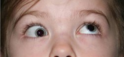
Patients, or in congenital cases more often a parent, note limited elevation of the affected eye specifically when it is turned toward the nose, sometimes with an audible or palpable click as the eye moves through the restricted range, along with a downward deviation (hypotropia) of the affected eye noticeable in the position of gaze up-and-in.
A compensatory head posture — chin elevation and/or a head tilt away from the affected side — is common, adopted to minimize the functional impact of the restriction and to help maintain binocular single vision in a comfortable head position.
Exam Findings
- Restricted elevation of the affected eye specifically in adduction, with elevation typically improving as the eye is moved into abduction — this gaze-dependent pattern (worse in adduction, better in abduction) is a key distinguishing feature
- A positive forced duction test — passive attempted elevation of the eye in adduction meets mechanical resistance, confirming a true restrictive process rather than a paretic one, and directly distinguishing Brown syndrome from inferior oblique palsy, which would show no such resistance on forced duction testing
- Widening of the palpebral fissure on attempted adduction-elevation, sometimes seen as the globe is mechanically prevented from elevating normally
- In trochleitis specifically, tenderness to palpation over the trochlea (superonasal orbital rim), supporting an inflammatory rather than purely structural mechanism
- Downshoot of the affected eye in attempted adduction, sometimes prominent enough to be a chief presenting concern
Differential Diagnosis
- Inferior oblique palsy — a paretic (not restrictive) limitation of elevation in adduction, distinguished by a negative forced duction test, since there is no mechanical restriction to overcome passively
- Superior oblique overaction — excessive, rather than restricted, depression in adduction, a fundamentally different direction of abnormal movement
- Double elevator palsy (monocular elevation deficiency) — restricted elevation in all positions of gaze, not specifically confined to the adducted position the way Brown syndrome’s restriction is
- Orbital floor fracture with inferior rectus entrapment — restricted elevation from a different mechanical cause, distinguished by trauma history, orbital imaging findings, and a different pattern of restriction on forced duction testing across gaze positions
Management
Many cases of congenital Brown syndrome are mild and stable, causing minimal functional impact, and can be observed when the head posture is comfortable and binocular vision is well maintained without treatment, with periodic reassessment rather than a single initial evaluation.
Trochleitis-related acquired Brown syndrome often responds well to anti-inflammatory treatment — oral or, in some cases, local corticosteroid injection near the trochlea — reflecting the reversible, inflammatory rather than fixed, structural nature of this specific subtype.
Surgical intervention — tenotomy or tendon-lengthening procedures at the superior oblique tendon or trochlea — is reserved for cases with a significant, bothersome head posture, significant hypotropia in primary position, or diplopia not adequately managed by observation or medical treatment, and is generally approached cautiously given the risk of inducing an iatrogenic superior oblique palsy if the tendon is weakened too aggressively during the procedure.
Follow-up after either medical or surgical treatment focuses on confirming that the restrictive pattern has genuinely resolved rather than simply becoming less symptomatic, since a persistently positive forced duction test can signal incomplete correction even when the head posture has improved.
Recurrence of trochleitis-related disease is possible, particularly when an underlying systemic inflammatory condition is present, so a return of symptoms after apparent resolution should prompt reassessment rather than assuming the initial diagnosis was mistaken.


Document what you see
Two smartphone imaging tools built for everyday clinic use — one for the slit lamp, one for the fundus.
From Choroida — the team behind this siteReferences
- Wright KW. Brown’s syndrome: diagnosis and management. Transactions of the American Ophthalmological Society.
- Kaban TJ, Smith K, Orton RB, Noel LP. Idiopathic Brown’s syndrome. Canadian Journal of Ophthalmology.
- American Academy of Ophthalmology. Basic and Clinical Science Course, Section 6: Pediatric Ophthalmology and Strabismus.
Test yourself
A few questions straight from this article.
-
What mechanism produces the limited eye movement in Brown syndrome?
Brown syndrome is a restrictive strabismus: the tendon-trochlea complex cannot glide freely. The superior oblique muscle itself is neither weak nor paralysed. -
How does elevation of the affected eye vary with horizontal gaze in Brown syndrome?
This gaze-dependent pattern, worse in adduction and better in abduction, is a key distinguishing feature of Brown syndrome. -
Which finding separates Brown syndrome from inferior oblique palsy?
Passive elevation meets mechanical resistance in Brown syndrome, confirming restriction. Inferior oblique palsy is paretic, so forced duction testing is negative. -
What underlies congenital Brown syndrome?
Congenital cases reflect a developmental abnormality of the superior oblique tendon or trochlea, such as a tendon that is too short or a trochlea that blocks smooth gliding. -
Which examination finding supports trochleitis as the cause of acquired Brown syndrome?
Tenderness over the trochlea at the superonasal orbital rim supports an inflammatory rather than purely structural mechanism. -
Which compensatory head posture do patients with Brown syndrome commonly adopt?
Chin elevation and a tilt away from the affected side minimise the functional impact of the restriction and help maintain binocular single vision comfortably. -
Which pattern indicates double elevator palsy rather than Brown syndrome?
Monocular elevation deficiency restricts elevation across all gaze positions, whereas the Brown syndrome restriction is confined to the adducted position. -
How is trochleitis-related acquired Brown syndrome usually treated?
This subtype is inflammatory and reversible rather than fixed and structural, so it often responds well to oral anti-inflammatory treatment or local corticosteroid injection near the trochlea. -
Which risk makes surgery for Brown syndrome an option approached cautiously?
Tenotomy or tendon-lengthening can weaken the tendon too aggressively and create a superior oblique palsy, so surgery is reserved for bothersome head posture, primary-position hypotropia or diplopia. -
Symptoms return months after successful treatment of trochleitis-related Brown syndrome. What does this most likely represent?
Trochleitis-related disease can recur, especially with an underlying systemic inflammatory condition, so returning symptoms warrant reassessment rather than doubting the original diagnosis.
