Optic perineuritis is inflammation of the optic nerve sheath, while the nerve itself is largely spared.
It often mimics optic neuritis at first presentation.
However, its age profile, visual pattern and MRI appearance differ.
Early recognition also changes the workup and the steroid plan.
What Optic Perineuritis Is
The inflammation targets the dura and arachnoid around the intraorbital optic nerve.
Many authors place it within the spectrum of orbital inflammatory disease.
Because the axons lie inside the inflamed sheath, they often escape severe early damage.
As a result, central vision can stay surprisingly good.
Who Gets Optic Perineuritis
Patients tend to be older than the typical optic neuritis population.
Purvin and colleagues described a wide age range, with many patients beyond middle age.
Therefore, an older adult labeled with “optic neuritis” deserves a second look.
All-fit Slit-Lamp Adapter
Record and share exactly what you see at the slit lamp. One adapter fits any slit lamp or surgical microscope — and any smartphone.
From Choroida — the team behind this siteSymptoms Patients Report
Pain is common and often prominent.
It usually worsens with eye movement and may precede any visual complaint.
Some patients describe blur or a dim patch off center.
Exam Findings in Optic Perineuritis
- Visual acuity normal or only mildly reduced
- Paracentral or arcuate field defects, with central sparing in some cases
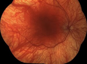
- Disc edema in a proportion of eyes, sometimes with peripapillary hemorrhage
- Mild or absent relative afferent pupillary defect
- Occasional orbital signs, such as mild proptosis or painful ocular motility
Disc Edema With Preserved Acuity
A swollen disc with good acuity usually points toward papilledema.
However, optic perineuritis can produce the same picture in one or both eyes.
Here the swelling reflects local sheath inflammation, and intracranial pressure stays normal.
Then a lumbar puncture, when needed, typically shows a normal opening pressure (see disc swelling causes).
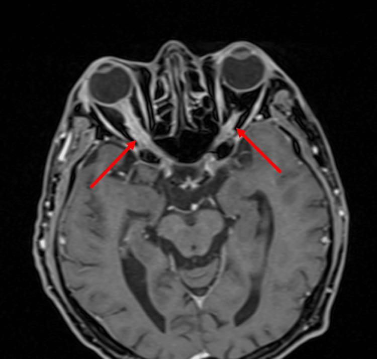
The Tram-Track MRI Sign in Optic Perineuritis
Contrast-enhanced, fat-suppressed orbital MRI is the key test.
The sheath enhances circumferentially while the nerve stays dark.
On axial images, this pattern creates two parallel lines, the “tram-track” sign.
On coronal images, the same ring looks like a doughnut around the nerve.
Optic Perineuritis Versus Optic Neuritis
Optic neuritis usually affects younger adults and causes central loss.
In contrast, optic perineuritis often spares the center of the field.
On MRI, optic neuritis enhances the nerve itself.
Also, perineuritis recovers less often without treatment and depends more on steroids.
Other Tram-Track Mimics
Optic nerve sheath meningioma also shows tram-track enhancement (see optic nerve sheath meningioma).
However, meningioma causes slow, painless loss and may calcify on CT.
Sarcoidosis, lymphoma, leukemic infiltration and metastasis can also involve the sheath.
Therefore, atypical features or a poor steroid response call for a biopsy discussion.
Causes of Optic Perineuritis
Many cases remain idiopathic.
However, secondary causes appear often enough to search for them every time.
Reported associations include syphilis, sarcoidosis, giant cell arteritis and granulomatosis with polyangiitis.
IgG4-related disease, tuberculosis, herpes zoster and Crohn disease also appear in case reports.
More recently, some patients with MOG antibody disease have shown perineural enhancement (see MOG optic neuritis).
Workup Before Treatment
First, order syphilis serology, because steroids alone may worsen an untreated infection.
Next, check ESR and CRP in older patients to screen for giant cell arteritis.
Then consider ACE, chest imaging, ANCA, IgG4 levels and MOG antibodies according to the history.
Steroid Response in Optic Perineuritis
Idiopathic disease usually responds quickly to corticosteroids.
Pain often eases within days, and vision tends to improve.
Indeed, a brisk response supports the diagnosis.
However, a poor response should prompt a search for infection, tumor or systemic inflammation.
How to Taper
Relapse often follows a rapid taper.
Therefore, most clinicians reduce the dose slowly, often over several months.
Patients with recurrences may need steroid-sparing agents.
When systemic disease is present, coordinate care with rheumatology.
Steroid Risks and Counseling
Explain that steroids control inflammation but cannot reverse established axonal loss.
Discuss sleep disturbance, mood change, hyperglycemia, raised blood pressure and bone loss.
Also warn about steroid-induced intraocular pressure rise and cataract with long courses.
Then agree on a clear follow-up schedule before the taper starts.
Prognosis of Optic Perineuritis
Most idiopathic patients regain good vision with timely treatment.
Delayed treatment, however, may leave permanent field loss.
Later, some discs turn pale as edema resolves.
Follow-Up Plan
Repeat acuity, color vision, pupils and visual fields at each visit.
Next, use OCT of the nerve fiber layer to track edema resolution and late thinning.
Finally, repeat MRI if symptoms recur or the course looks atypical.
Pitfalls to Avoid
- Do not label every painful swollen disc in an older adult as optic neuritis
- Never start steroids before ordering syphilis serology
- Avoid a rapid taper in idiopathic disease
- Do not assume tram-track enhancement always means meningioma
- Never miss giant cell arteritis in patients over 50


Document what you see
Two smartphone imaging tools built for everyday clinic use — one for the slit lamp, one for the fundus.
From Choroida — the team behind this siteReferences
- Purvin V, Kawasaki A, Jacobson DM. Optic perineuritis: clinical and radiographic features. Arch Ophthalmol. 2001;119:1299-1306.
- Hickman SJ. Optic perineuritis. Curr Neurol Neurosci Rep. 2016;16:16.
- American Academy of Ophthalmology. Basic and Clinical Science Course, Section 5: Neuro-Ophthalmology. San Francisco: AAO; 2023.
- Salmon JF. Kanski’s Clinical Ophthalmology: A Systematic Approach. 9th ed. Elsevier; 2020.
- Miller NR, Newman NJ, Biousse V, Kerrison JB. Walsh and Hoyt’s Clinical Neuro-Ophthalmology. 6th ed. Lippincott Williams and Wilkins; 2005.

Writes and reviews the clinical articles on Educate. Every post is checked against peer-reviewed sources before it goes live.