
Optic nerve sheath meningioma (ONSM) is a benign tumor that arises from the arachnoid cap cells of the meninges that surround the optic nerve.
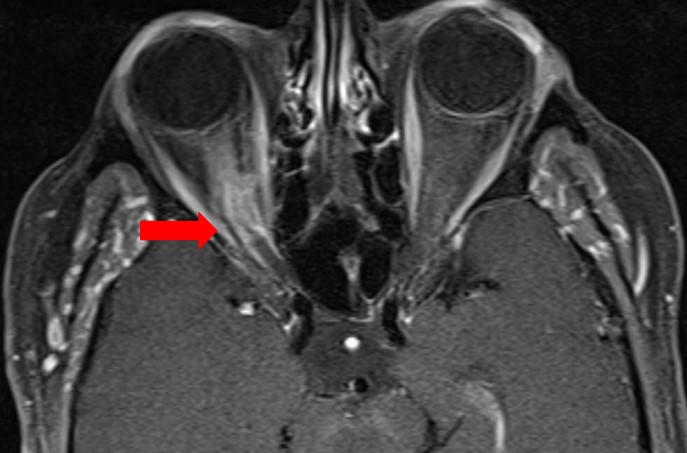
It typically affects middle-aged women, and it presents with slowly progressive, painless loss of vision.
The tumor grows along the nerve and compresses it from the outside, and treatment must balance control of the tumor with preservation of the vision that remains.
What Is an Optic Nerve Sheath Meningioma?
ONSM accounts for a small fraction of all meningiomas and for a notable share of primary optic nerve tumors.
It can arise within the orbit or extend into the orbit from the intracranial portion of the nerve, and it may spread through the optic canal toward the chiasm.
Most cases are unilateral, and bilateral tumors suggest neurofibromatosis type 2, particularly in a younger patient.
Hormonal influences have been proposed because of the female predominance, but the cause is not established.
Clinical Features
The classic triad is progressive visual loss, optic atrophy, and optociliary shunt vessels, although all three are rarely present at the first visit (see optociliary shunt vessels).
- Painless, gradual loss of acuity and visual field, often with a relative afferent pupillary defect
- Disc swelling in the early phase, followed by pallor
- Shunt vessels between the retinal and choroidal circulations at the disc
- Mild proptosis, sometimes with restricted motility and diplopia
- Transient visual obscurations in some patients
Visual field defects may be peripheral constriction or central scotoma, and they vary with the site of compression.
Fundus Explorer Pro
Photograph the retinal findings described here with the phone already in your pocket — 22 D optics and built-in illumination in one handheld unit.
From Choroida — the team behind this siteImaging
MRI with fat suppression and contrast shows a thick, enhancing sheath that surrounds a nonenhancing nerve, which produces the “tram-track” sign on axial images.
CT may show linear or granular calcification of the sheath, and it also shows hyperostosis of the adjacent bone when the tumor extends.
The imaging is often diagnostic, and biopsy is avoided because it can damage the nerve and the blood supply.
Thin sections through the optic canal and the cavernous sinus show intracranial spread.
Differential Diagnosis
- Optic nerve glioma: fusiform enlargement of the nerve itself, mostly in children (see optic nerve glioma)
- Optic perineuritis: inflammation of the sheath that mimics the tram-track sign, with pain and a response to steroids
- Sarcoidosis, IgG4-related disease, and lymphoma: infiltrative causes that may need biopsy
- Idiopathic orbital inflammation: a painful, acute course
- Papilledema or compressive neuropathy from other lesions: a different pattern on imaging
Management
Observation is appropriate when vision is good and the tumor is small, with serial imaging and visual testing.
Fractionated stereotactic radiotherapy is the treatment of choice for tumors that threaten vision, and it often stabilizes or improves visual function.
Surgical removal of the tumor is rarely indicated for the intraorbital part, because it frequently sacrifices vision, and it is reserved for tumors that extend toward the chiasm, or for a blind, disfiguring eye.
Long-term follow-up is needed, because radiation effects such as retinopathy and optic neuropathy can occur after months or years.
Patients should be counselled that the goal is to preserve what remains of the vision, and that some visual loss may not recover.
The Visual Course Without Treatment
Left untreated, vision declines slowly, over years, and the rate varies from patient to patient.
Some patients remain stable for a long period, and others lose vision more quickly, particularly when the tumor extends through the canal.
The tumor often causes a mild proptosis, and extraocular motility may become restricted late in the disease.
A growing tumor may involve the chiasm and the other optic nerve, so imaging at intervals is important.
Why Radiotherapy Is Preferred
Studies of long-term outcome show that fractionated stereotactic radiotherapy stabilizes or improves vision in most patients, while observation alone leads to progressive loss in many and surgery alone carries a high rate of vision loss.
The radiation dose is delivered in small fractions to limit damage to the nerve and the retina, and modern planning techniques focus the dose on the tumor.
Patients should be told that improvement is possible, but that complete recovery is uncommon, and that visual function should be monitored for years after treatment.
Special Situations
In young patients, bilateral disease, or tumors with multiple lesions, suspect neurofibromatosis type 2 and refer for genetic assessment.
In pregnancy, the tumor may enlarge because of hormonal influences, and management is individualized with the obstetric and neurosurgical teams.
A tumor with intracranial extension needs neurosurgical evaluation, and a combined approach may be required.
Patients should be offered support for living with visual loss, since many are in their working years and have family responsibilities.


Document what you see
Two smartphone imaging tools built for everyday clinic use — one for the slit lamp, one for the fundus.
From Choroida — the team behind this siteReferences
- Dutton JJ. Optic nerve sheath meningiomas. Surv Ophthalmol. 1992;37:167-183.
- Turbin RE, Thompson CR, Kennerdell JS, et al. A long-term visual outcome comparison in patients with optic nerve sheath meningioma managed with observation, surgery, radiotherapy, or surgery and radiotherapy. Ophthalmology. 2002;109:890-899.
- Liu GT, Volpe NJ, Galetta SL. Neuro-Ophthalmology: Diagnosis and Management. 3rd ed. Elsevier; 2019.
- American Academy of Ophthalmology. Basic and Clinical Science Course, Section 7: Oculofacial Plastic and Orbital Surgery. AAO; current edition.

Writes and reviews the clinical articles on Educate. Every post is checked against peer-reviewed sources before it goes live.


