Ocular albinism is an inherited hypopigmentation that mainly affects the eye, while skin and hair look near normal.
The classic form is X-linked ocular albinism type 1, also called Nettleship-Falls albinism.
Affected boys present with nystagmus, reduced vision and photophobia, so the diagnosis often starts at the slit lamp.
Genetics of X-Linked Ocular Albinism
Mutations in the GPR143 gene on Xp22 cause the X-linked form.
This gene encodes a G protein-coupled receptor on the melanosome membrane.
When it fails, melanosomes grow abnormally large and pigmentation becomes disordered.
As a result, the iris and retinal pigment epithelium carry too little effective melanin.
How Ocular Albinism Differs From Oculocutaneous Forms
Oculocutaneous albinism (OCA) is autosomal recessive and lightens skin and hair as well as the eyes.
In contrast, ocular albinism spares visible skin pigment in most patients.
Both groups share a similar eye phenotype, therefore pedigree, skin color and genetic testing decide the label.
Many patients once labeled autosomal recessive ocular albinism later proved to have mild OCA.
Fundus Explorer Pro
Photograph the retinal findings described here with the phone already in your pocket — 22 D optics and built-in illumination in one handheld unit.
From Choroida — the team behind this sitePresenting Symptoms
Nystagmus usually appears in the first months of life.
Parents then notice that the infant squints in bright light.
Acuity is moderately reduced, although the range between patients is wide.
Strabismus and high astigmatism are also common.
Iris Transillumination in Ocular Albinism
Retroillumination at the slit lamp is the quickest office test.
Direct a narrow beam through the pupil and watch the red reflex glow back through the iris.
Affected males often transilluminate diffusely, while milder cases show punctate or radial defects.
Several other conditions produce the same sign (see iris transillumination defects).
Fundus Findings in Affected Males
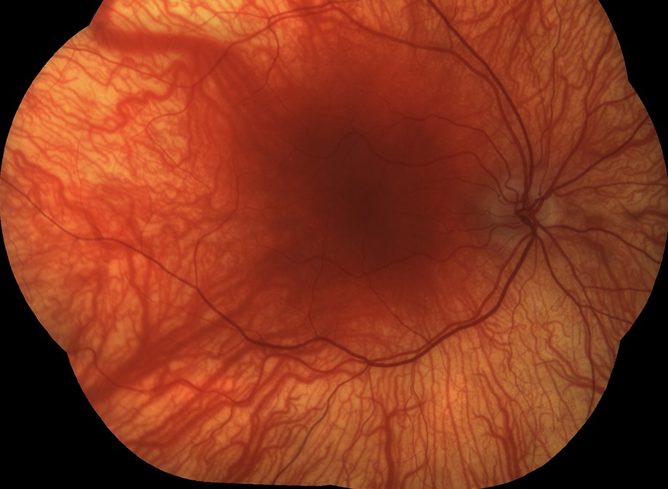
On dilated exam, the fundus looks pale, and large choroidal vessels stand out against the thin pigment layer.
The macula lacks its normal darker pigment, and the foveal reflex is absent.
In addition, retinal vessels may course close to the presumed foveal center.
Finally, the optic disc may appear small or gray.
Foveal Hypoplasia on OCT
OCT shows inner retinal layers persisting across the fovea.
The foveal pit is shallow or absent.
Thomas and colleagues graded these changes, and higher grades predicted poorer acuity.
Our post on foveal hypoplasia in albinism covers the imaging detail.
Optic Pathway Misrouting
In albinism, excess temporal retinal fibers cross at the chiasm.
Multichannel visual evoked potentials detect this crossed asymmetry.
Therefore, a VEP can support the diagnosis when ocular signs look subtle.
However, recordings in young infants need pediatric expertise.
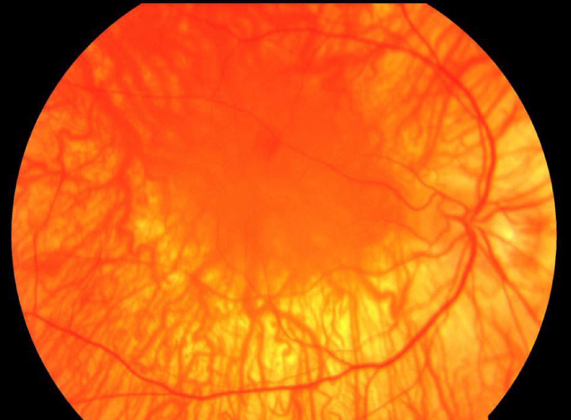
The Mud-Splatter Carrier Fundus
Female carriers usually see normally and have no nystagmus.
However, the peripheral fundus often shows a mosaic of pigmented and depigmented patches.
Clinicians call it the mud-splatter pattern.
It reflects random X inactivation, or lyonization, within the retinal pigment epithelium.
Iris Signs in Ocular Albinism Carriers
Carriers can also show patchy or radial iris transillumination.
Charles and colleagues reported that careful iris and fundus examination detected changes in many obligate carriers.
Similar mosaic patterns occur in other X-linked retinal diseases (see RPGR carrier signs).
Diagnostic Workup for Ocular Albinism
First, document nystagmus, refraction and alignment.
Next, perform retroillumination and a dilated fundus exam.
Then obtain macular OCT if the child cooperates.
Finally, examine the mother, because carrier signs support X-linked inheritance.
GPR143 testing can confirm the diagnosis in many cases.
Differential Diagnosis
- Oculocutaneous albinism, with skin and hair hypopigmentation
- Hermansky-Pudlak syndrome, with bleeding tendency
- Chediak-Higashi syndrome, with recurrent infections
- Aniridia and other PAX6 disorders
- Idiopathic infantile nystagmus, with a normal fovea
Systemic Red Flags
Ocular albinism itself carries no systemic disease.
However, easy bruising, nosebleeds or frequent infections suggest syndromic albinism.
In those children, involve hematology before any surgery.
Hermansky-Pudlak syndrome can also cause pulmonary fibrosis later in life.
Managing Ocular Albinism
No treatment restores melanin or rebuilds the fovea.
Therefore, care aims to maximize the vision the child already has.
Full cycloplegic refraction comes first, because uncorrected astigmatism further degrades acuity.
Tinted lenses reduce glare, while low vision aids support schoolwork.
Also, some children adopt a head posture that uses the nystagmus null point.
Counseling Families About Ocular Albinism
Each son of a carrier mother has a 50 percent risk of being affected.
Each daughter has a 50 percent chance of being a carrier.
An affected man passes the mutation to all daughters and no sons.
Also, explain that vision usually stays stable through life.
Pitfalls to Avoid
- Do not miss transillumination in dark irides, so dim the room first
- Never diagnose idiopathic nystagmus before checking the fovea
- Do not skip the mother’s fundus examination
- Never assume normal skin excludes albinism


Document what you see
Two smartphone imaging tools built for everyday clinic use — one for the slit lamp, one for the fundus.
From Choroida — the team behind this siteReferences
- Bassi MT, Schiaffino MV, Renieri A, et al. Cloning of the gene for ocular albinism type 1 from the distal short arm of the X chromosome. Nat Genet. 1995;10:13-19.
- Charles SJ, Moore AT, Grant JW, Yates JR. Genetic counselling in X-linked ocular albinism: clinical features of the carrier state. Eye. 1992;6:75-79.
- Thomas MG, Kumar A, Mohammad S, et al. Structural grading of foveal hypoplasia using spectral-domain optical coherence tomography: a predictor of visual acuity? Ophthalmology. 2011;118:1653-1660.
- Summers CG. Albinism: classification, clinical characteristics, and recent findings. Optom Vis Sci. 2009;86:659-662.
- Bowling B. Kanski’s Clinical Ophthalmology: A Systematic Approach. 8th ed. Elsevier; 2016.

Writes and reviews the clinical articles on Educate. Every post is checked against peer-reviewed sources before it goes live.