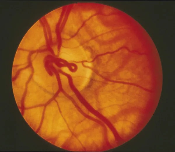
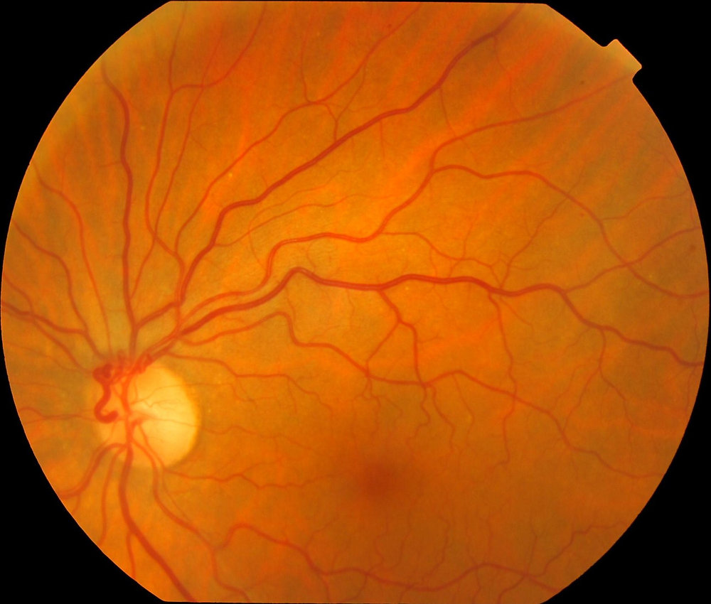
Prepapillary loop is a congenital variant in which a retinal vessel rises from the optic disc into the vitreous, then returns.
Most affected eyes remain asymptomatic for life.
However, rare occlusions and vitreous hemorrhage make accurate recognition useful.
What Is a Prepapillary Loop?
The vessel forms an arc, coil or figure-of-eight in front of the disc.
Blood then continues into a retinal branch that supplies a defined sector.
Most reported loops are arterial, while venous loops appear less often.
Therefore, the examiner should trace both limbs and identify the territory they supply.
How a Prepapillary Loop Develops
The usual explanation links the loop to fetal vascular development at the disc.
Developing retinal vessels may grow into Bergmeister papilla, the glial tissue surrounding the hyaloid artery.
They then turn back toward the retina and leave a loop as that tissue regresses.
However, this sequence remains a developmental hypothesis, because direct proof is limited.
Fundus Explorer Pro
Photograph the retinal findings described here with the phone already in your pocket — 22 D optics and built-in illumination in one handheld unit.
From Choroida — the team behind this siteFundus Signs of a Prepapillary Loop
Stereoscopic examination provides the best view, because depth is the defining feature.
Look for these signs:
- A smooth, regular vessel rising from the disc surface
- A clear path back to the disc or nearby retina
- Caliber similar to a normal retinal vessel
- Possible pulsation or movement with eye motion
- A fine white sheath around the loop
Imaging a Prepapillary Loop
Color fundus photography documents shape, position and any change over time.
OCT through the disc can show the vessel rising above the retinal surface into the vitreous.
In addition, OCT angiography may display flow, although projection and segmentation artifacts can limit interpretation.
Serial photographs help most, because a congenital loop should keep a stable configuration.
Branch Retinal Artery Occlusion
Arterial loops have a recognized association with branch retinal artery occlusion in the supplied territory.
Proposed mechanisms include turbulent flow, kinking, torsion and thrombosis, although causation is hard to prove in individual cases.
Therefore, the workup should still include carotid, cardiac and inflammatory causes when clinically appropriate.
Patients may also describe transient episodes before a fixed defect appears.
Vitreous Hemorrhage and Other Bleeding
The elevated vessel extends into the vitreous, so vitreous traction may theoretically injure it.
Case reports describe vitreous hemorrhage, sometimes recurrent, in eyes with these loops.
Hyphema has also appeared in reports, although it is very rare.
Nevertheless, a hemorrhage still requires a dilated search for retinal tears and neovascularization.
Prepapillary Loop vs Disc Neovascularization
Neovascularization of the disc forms fine, irregular, branching networks that usually leak on angiography.
It also follows ischemic disease, such as proliferative diabetic retinopathy or retinal vein occlusion.
A congenital loop instead shows a single smooth vessel with normal caliber.
Then, examine the retinal periphery, because ischemic findings support an acquired process.
Prepapillary Loop vs Optociliary Shunt
Optociliary shunts are acquired collaterals that divert retinal venous blood toward the choroidal circulation.
They usually lie on the disc surface and do not project into the vitreous.
Moreover, they suggest chronic venous obstruction, such as prior vein occlusion or optic nerve sheath meningioma (see optociliary shunt vessels).
Also, a persistent hyaloid artery runs toward the lens and has no perfused return limb.
Managing an Asymptomatic Prepapillary Loop
An asymptomatic, typical loop requires no treatment.
No evidence supports prophylactic laser, antiplatelet therapy or vitrectomy for the isolated finding.
Instead, document the lesion with a photograph and record the supplied quadrant.
Routine eye examinations are generally sufficient, although follow-up intervals should reflect other ocular findings.
Treating Complications of a Prepapillary Loop
Treatment targets the complication, because the loop itself rarely needs intervention.
An acute artery occlusion calls for urgent systemic vascular evaluation, even with a visible local anomaly.
Vitreous hemorrhage often allows observation, provided ultrasound excludes retinal detachment when the view is poor.
Finally, vitrectomy can clear nonresolving or recurrent hemorrhage, but it carries surgical risks, including cataract and retinal tears.
Counseling Patients with a Prepapillary Loop
Explain that the patient was born with this vessel pattern and that it is not a tumor.
Also, state that complications are rare and that the available evidence cannot predict individual risk.
Advise urgent review for sudden vision loss, a new field defect, or a shower of floaters.
A copy of the fundus photograph can also help future examiners.
Pitfalls to Avoid
- Do not label every vessel on the disc as congenital
- Do not treat a smooth, stable loop as neovascularization
- Never skip a systemic workup after arterial occlusion
- Do not rely on a nondilated, monocular view


Document what you see
Two smartphone imaging tools built for everyday clinic use — one for the slit lamp, one for the fundus.
From Choroida — the team behind this siteReferences
- Degenhart W, Brown GC, Augsburger JJ, Magargal L. Prepapillary vascular loops. Ophthalmology. 1981;88:1126-1131.
- Brown GC, Magargal LE, Augsburger JJ, Shields JA. Preretinal arterial loops and retinal arterial occlusion. Am J Ophthalmol. 1979;87:646-651.
- Shakin EP, Shields JA, Augsburger JJ, Brown GC. Clinicopathologic correlation of a prepapillary vascular loop. Retina. 1988;8:55-58.
- Strassman IB, Desai UR. Prepapillary vascular loop and a recurrent vitreous hemorrhage. Retina. 1997;17:166-167.
- American Academy of Ophthalmology. Basic and Clinical Science Course, Section 12: Retina and Vitreous. San Francisco: AAO.
Test yourself
A few questions straight from this article.
-
What is a prepapillary vascular loop?
A prepapillary loop is a congenital variant in which a retinal vessel rises from the disc into the vitreous as an arc, coil or figure-of-eight, then returns. -
Which vessel type forms most reported prepapillary loops?
Most reported prepapillary loops are arterial, with venous loops less often; both limbs should be traced to identify the supplied territory. -
Into which fetal structure are developing retinal vessels thought to grow to form a prepapillary loop?
The usual hypothesis is that retinal vessels grow into Bergmeister papilla, the glial tissue around the hyaloid artery, and leave a loop as it regresses; direct proof is limited. -
Why does stereoscopic examination give the best view of a prepapillary loop?
Because the loop projects forward from the disc, depth is its defining feature, so a nondilated monocular view should not be relied on. -
On serial fundus photographs, what behaviour is expected of a congenital prepapillary loop?
Serial photographs help most because a congenital loop should keep a stable configuration; OCT can also show the vessel rising into the vitreous. -
Which occlusion has a recognized association with arterial prepapillary loops?
Arterial loops are associated with branch retinal artery occlusion in their territory, with turbulent flow, kinking, torsion and thrombosis proposed as mechanisms. -
A patient with a known prepapillary loop develops an acute branch retinal artery occlusion. What does management require?
Causation is hard to prove in individual cases, so carotid, cardiac and inflammatory causes are still evaluated urgently despite the visible local anomaly. -
Which feature distinguishes a prepapillary loop from neovascularization of the disc?
Disc neovascularization forms fine, irregular, branching networks that usually leak and follow ischemia; a congenital loop is a single smooth vessel of normal caliber. -
How does an optociliary shunt differ from a prepapillary loop?
Optociliary shunts are acquired venous collaterals to the choroidal circulation that lie on the disc surface, suggesting chronic venous obstruction such as meningioma. -
What is the appropriate management of an asymptomatic, typical prepapillary loop?
No evidence supports prophylactic laser, antiplatelets or vitrectomy; the loop is documented photographically and routine examinations usually suffice.

Writes and reviews the clinical articles on Educate. Every post is checked against peer-reviewed sources before it goes live.