
LASIK flap complications arise during creation of the corneal flap, in the early postoperative period, or years later after trauma.
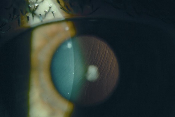
Most are uncommon with modern microkeratomes and femtosecond lasers, but they can cause lasting visual disturbance if clinicians miss or mishandle them.
Knowing the pattern of each complication helps the surgeon to react in the right way.
Intraoperative LASIK Flap Complications
- Incomplete flap: the microkeratome stops before the flap is complete, and the laser should not be applied
- Thin flap and buttonhole: a hole in the flap, usually over a steep cornea, and the ablation should also be postponed
- Free cap: the flap is fully detached, and it must be saved and repositioned with correct orientation
- Irregular or decentered flap: from poor suction or a poor cornea
- Epithelial defect: more common in older patients and in those with basement membrane disease (see map-dot-fingerprint dystrophy)
- Suction loss: interrupts the cut, and the procedure should be stopped
- Opaque bubble layer with femtosecond lasers, which blocks tracking
The surgeon should stop and reschedule the ablation when the flap is irregular, since going ahead with a defective flap leads to irregular astigmatism.
LASIK Flap Complications: Early Postoperative Problems
- Flap striae or folds: wrinkles in Bowman layer that can cause irregular astigmatism and ghosting
- Flap slippage or displacement: from rubbing or trauma in the first hours and days
- Diffuse lamellar keratitis: a sterile inflammation in the interface (see diffuse lamellar keratitis)
- Infectious keratitis: including bacterial and atypical mycobacterial infection (see nontuberculous mycobacterial keratitis)
- Epithelial ingrowth: cells growing beneath the flap edge (see epithelial ingrowth)
- Interface debris and pressure-induced stromal keratitis
All-fit Slit-Lamp Adapter
Record and share exactly what you see at the slit lamp. One adapter fits any slit lamp or surgical microscope — and any smartphone.
From Choroida — the team behind this siteLate Problems
Flaps remain only weakly attached to the stroma for life, and traumatic dislocation can occur years after surgery.
Late-onset striae, epithelial ingrowth, and ectasia are also other long-term problems (see post-LASIK ectasia).
Advise patients that blunt trauma to the eye, even years later, can displace the flap, and that sports require eye protection.
Evaluation for LASIK Flap Complications
Examine the flap at the slit lamp with a narrow beam, using retroillumination and fluorescein to show striae as negative staining lines.
In addition, check the interface for cells, debris, or epithelium, and examine the edges for gaps.
Topography and refraction demonstrate irregular astigmatism, and OCT can show flap thickness and interface changes (see anterior segment OCT).
Management for LASIK Flap Complications
- Striae: lift the flap, rehydrate or stretch it, smooth it with a spatula, and reposition it, and in persistent cases add sutures
- Displaced flap: reposition it urgently, clean the interface, and scrape epithelium from the bed if it has grown in
- Buttonhole or free cap: replace the tissue carefully, allow healing for months, and consider surface ablation later
- Diffuse lamellar keratitis: frequent topical steroids and, in severe cases, interface irrigation
- Infection: culture, lift the flap, irrigate, and start intensive antibiotics
- Epithelial ingrowth: observe if peripheral and stable, and remove if it threatens vision or melts the flap
The decision on timing is important, because early intervention for striae and dislocation leads to better results.
Prevention
Careful patient selection, accurate suction, a good surgical technique, and a clean environment reduce complication rates.
Patients should also receive clear postoperative instructions about not rubbing the eyes, about eye shields, and about reporting symptoms.
Managing Specific Problems in More Detail
For flap striae, the best time to intervene is within the first days, when the epithelium has not healed over the fold lines.
Lift the flap under a microscope, hydrate the bed, and stretch the flap in the direction perpendicular to the fold with a moist sponge or spatula, then allow it to adhere.
Some persistent striae require suturing, or removal of the epithelium over the fold to flatten it.
A bandage lens may help.
For a free cap, mark the epithelial side with a stain before removal, keep the tissue moist on a dry surface in a chamber, and replace it with the epithelium up after the ablation or after postponing it.
Interface Inflammation Versus Infection
Early after LASIK, diffuse lamellar keratitis appears within a day or two as sand-like granules in the interface.
It responds to topical steroids.
Infection tends to appear later, with focal infiltrates, pain, and progressive worsening, and delayed onset after several days suggests atypical mycobacteria or fungi.
When in doubt, lift the flap, culture the interface, and treat for infection while the results are pending.
Advice for Patients
Tell patients to avoid rubbing the eyes, to wear the shield at night for the recommended time, and to avoid swimming and dusty environments in the first weeks.
Explain that flap displacement can occur from trauma, and that any sudden blur, pain, or foreign body sensation requires urgent review.
Doctors recognize contact sports and airbag deployments as causes of late flap dislocation.
A note in the record helps future clinicians to be aware of the history of LASIK.
For the latest studies, see the PubMed results for LASIK flap complications.


Document what you see
Two smartphone imaging tools built for everyday clinic use — one for the slit lamp, one for the fundus.
From Choroida — the team behind this siteReferences
- Melki SA, Azar DT. LASIK complications: etiology, management, and prevention. Surv Ophthalmol. 2001;46:95-116.
- Stulting RD, Carr JD, Thompson KP, et al. Complications of laser in situ keratomileusis for the correction of myopia. Ophthalmology. 1999;106:13-20.
- Ambrósio R Jr, Wilson S. LASIK vs LASEK vs PRK: advantages and indications. Semin Ophthalmol. 2003;18:2-10.
- American Academy of Ophthalmology. Basic and Clinical Science Course, Section 13: Refractive Surgery. AAO; current edition.

Writes and reviews the clinical articles on Educate. Every post is checked against peer-reviewed sources before it goes live.


