Fluorescein angiography patterns fall into two groups: hyperfluorescence and hypofluorescence.
Each group has a few clear causes.
Therefore, a systematic approach turns a confusing picture into a short list of diagnoses.
How Fluorescein Angiography Patterns Arise
Sodium fluorescein is injected into an arm vein.
It binds mostly to albumin, and the free fraction fluoresces under blue light.
Also, the dye leaves the choroidal capillaries freely but not the retinal vessels.
As a result, leakage patterns separate the two circulations.
Phases and Fluorescein Angiography Patterns
The choroidal flush appears first, about 10 to 15 seconds after injection.
The arterial, arteriovenous and venous phases follow within the first minute.
Also, late frames at 5 to 10 minutes show staining and leakage.
Therefore, timing helps to interpret each finding.
Fundus Explorer Pro
Photograph the retinal findings described here with the phone already in your pocket — 22 D optics and built-in illumination in one handheld unit.
From Choroida — the team behind this siteNormal Fluorescein Angiography Patterns
The dye fills the retinal arteries and then the veins with laminar flow.
The fovea looks dark because of xanthophyll and a thick pigment layer.
Also, the optic disc stains late.
As a result, a normal study sets the baseline for comparison.
Four Mechanisms of Hyperfluorescence
Hyperfluorescence can come from a window defect, pooling, staining or leakage.
A fifth cause is autofluorescence of structures such as optic disc drusen.
Also, each mechanism has a typical time course.
Therefore, ask which of them fits the pattern.
Window Defect
A window defect shows increased visibility of choroidal fluorescence through a thin or missing pigment epithelium.
It appears early, peaks with the choroidal flush and fades with it.
Also, its size and shape stay constant.
As a result, it suggests atrophy of the epithelium.
Pooling
Pooling is the collection of dye in a space under the retina or the pigment epithelium.
It fills early and stays bright with sharp borders.
Also, serous detachments and central serous disease show pooling.
Therefore, pooling indicates a defect in the outer barrier.
Staining
Staining is the late uptake of dye by solid tissue such as drusen or a scar.
It appears gradually and persists without a change in size.
Also, the dye washes out slowly.
As a result, staining marks static lesions.
Leakage
Leakage is the escape of dye from abnormal vessels into the retina or subretinal space.
It grows in size and brightness over time with blurred borders.
Also, macular edema, neovascularization and choroidal neovascularization all leak.
Therefore, increasing hyperfluorescence points to active disease.
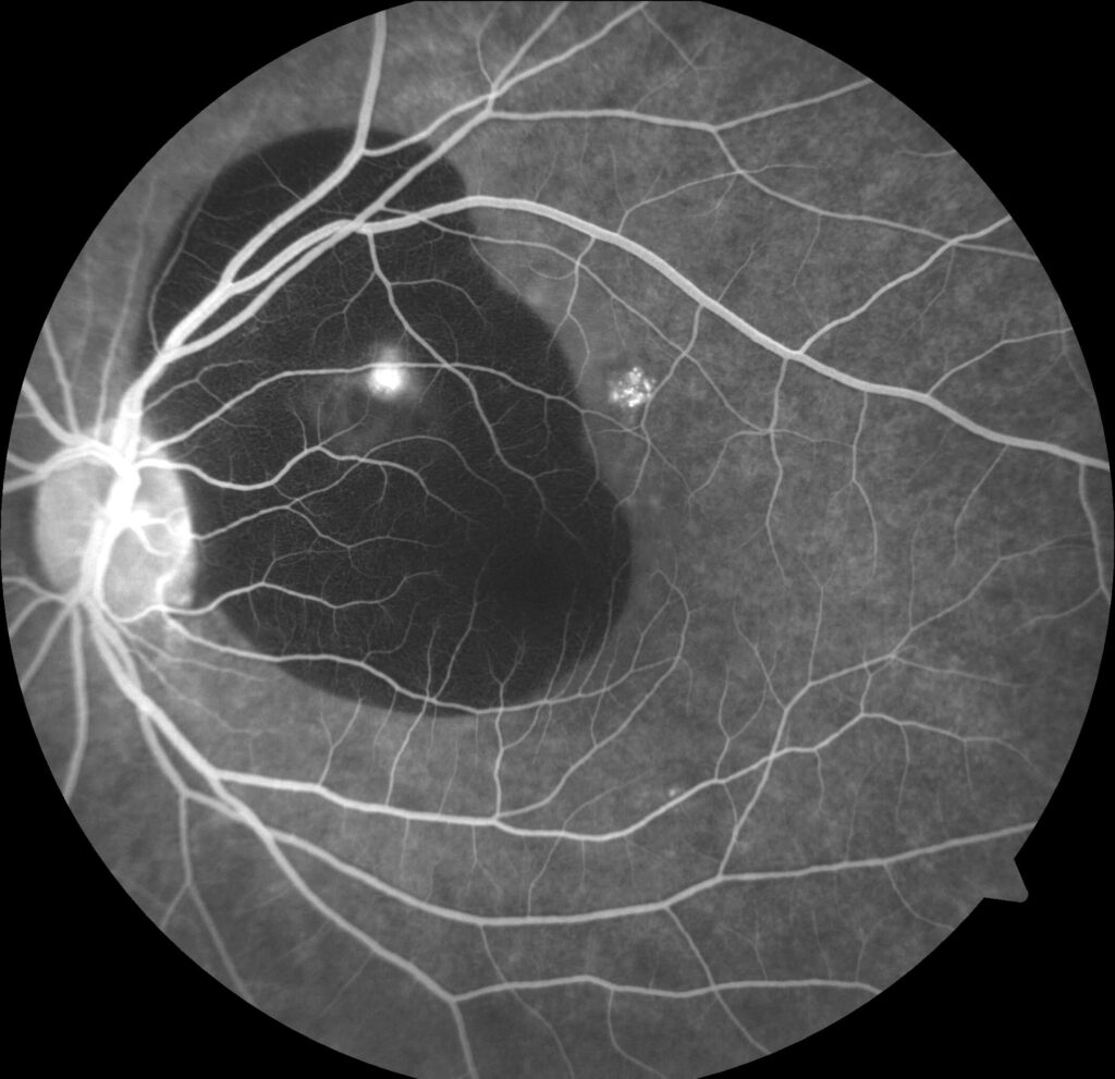
Fluorescein Angiography Patterns of Leakage
Cystoid edema shows a petal-like pattern in the late frames.
Classic neovascularization shows a lacy early network and late pooling.
Also, occult lesions show stippled late leakage without a clear source.
In addition, the pattern guides treatment (see diabetic macular edema).
Hypofluorescence from Blockage
Blockage hides normal fluorescence behind blood, pigment or exudate.
The size and shape follow the blocking material.
Also, preretinal blood blocks both retinal and choroidal fluorescence, while deeper blood blocks only the choroid.
Therefore, the depth of the lesion can be inferred.
Hypofluorescence from Filling Defects
A filling defect means the vessels are not perfused.
Nonperfusion in vein occlusion or diabetes shows large dark areas.
Also, choroidal ischemia shows patchy filling defects.
As a result, a dark area with no vessels suggests ischemia.
Distinguishing Blockage from Filling Defects
Compare the dark area with the red-free and color images.
Blood or pigment explains blockage.
Also, a normal color image with a dark angiogram points to a filling defect.
Therefore, always review the photographs next to the angiogram.
Artifacts in Fluorescein Angiography Patterns
Poor media, a small pupil and motion blur reduce quality.
Pseudofluorescence from filter overlap can mimic early staining.
Also, nausea and a bright skin may reflect dye reactions.
In addition, check the timing label on each frame.
Adverse Reactions
Nausea and vomiting occur in a minority of patients.
Severe reactions are rare, and anaphylaxis occurs in a very small number.
Also, Yannuzzi and colleagues and Kwiterovich and colleagues reported these rates in large surveys.
Therefore, have emergency equipment on hand.
Combining Fluorescein Angiography Patterns With OCT
OCT shows structure and angiography shows function.
Together they reveal the mechanism of fluid and the activity of vessels.
Also, OCT angiography can replace some angiograms (see indocyanine green angiography).
As a result, use each test for the question it answers best.
Pitfalls to Avoid
- Do not call every bright area leakage without checking later frames
- Never skip the color photograph when interpreting a dark area
- Do not forget to note the timing of each pattern
- Never inject dye without screening for prior reactions
For the latest studies, see the PubMed results for fluorescein angiography patterns.


Document what you see
Two smartphone imaging tools built for everyday clinic use — one for the slit lamp, one for the fundus.
From Choroida — the team behind this siteReferences
- Yannuzzi LA, Rohrer KT, Tindel LJ, et al. Fluorescein angiography complication survey. Ophthalmology. 1986;93:611-617.
- Kwiterovich KA, Maguire MG, Murphy RP, et al. Frequency of adverse systemic reactions after fluorescein angiography: results of a prospective study. Ophthalmology. 1991;98:1139-1142.
- Marmor MF, Ravin JG. Fluorescein angiography: insight and serendipity a half century ago. Arch Ophthalmol. 2011;129:943-948.
- Gass JDM. Stereoscopic Atlas of Macular Diseases: Diagnosis and Treatment. 4th ed. Mosby; 1997.

Writes and reviews the clinical articles on Educate. Every post is checked against peer-reviewed sources before it goes live.