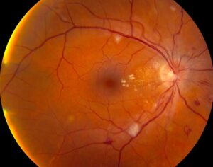
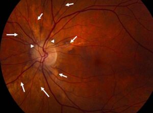
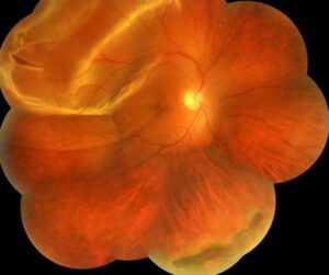
Retinoschisis vs detachment is a common peripheral retina question, because both conditions elevate the retina on fundus examination.
Degenerative retinoschisis splits the neurosensory retina, while rhegmatogenous detachment separates it from the retinal pigment epithelium (RPE).
Therefore, the plane of separation explains most clinical clues.
Anatomy of Retinoschisis vs Detachment
Typical degenerative schisis splits the retina at the outer plexiform layer.
The outer wall stays against the RPE.
A rhegmatogenous detachment, however, lifts the full neurosensory retina above subretinal fluid.
Symptoms and Risk Factors
Retinoschisis usually causes no symptoms, so it often appears during a routine dilated exam.
Detachment, in contrast, typically brings flashes, floaters and an advancing shadow.
However, a chronic inferior detachment can also remain silent.
Schisis favors older, hyperopic patients, while detachment is linked with myopia, lattice degeneration, prior cataract surgery and trauma.
Because these profiles overlap, risk factors only shift suspicion.
Fundus Explorer Pro
Photograph the retinal findings described here with the phone already in your pocket — 22 D optics and built-in illumination in one handheld unit.
From Choroida — the team behind this siteExam Signs in Retinoschisis vs Detachment
Schisis usually forms a smooth, immobile dome, most often inferotemporal.
Its thin inner wall may look transparent, with fine white dots and sheathed vessels.
A fresh detachment, by comparison, looks corrugated and moves with eye movement.
However, a chronic detachment can become thin and smooth, which makes it a convincing mimic.
Vitreous Pigment and Break Patterns
Next, examine the anterior vitreous at the slit lamp.
Pigment granules (Shafer sign) strongly suggest a retinal break.
Uncomplicated schisis should not produce this finding.
Scleral depression then lets you inspect both walls.
Inner wall holes tend to be small and round, while outer wall breaks are larger with rolled margins.
Signs of Chronicity
A pigment demarcation line suggests a present or past detachment component.
Subretinal strands, macrocysts and RPE atrophy also point toward long-standing detachment.
Therefore, any of them should prompt a search for subretinal fluid.
Visual Field in Retinoschisis vs Detachment
Schisis classically gives an absolute scotoma with steep borders, because the split interrupts neural transmission.
Detachment, however, typically causes a relative scotoma.
Still, far peripheral testing has limits, so use the field as supporting evidence.
Laser Uptake in Retinoschisis vs Detachment
A low-energy test burn can separate the two when the diagnosis stays uncertain.
The apposed outer wall of schisis takes up laser, and a visible burn follows.
Detached retina, however, shows little uptake because subretinal fluid separates it from the RPE.
This test differs from laser retinopexy, which seals a retinal break.
Since OCT answers the question noninvasively, few clinicians need this test now.
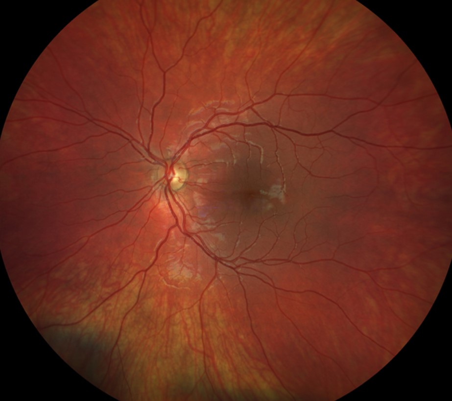
OCT in Retinoschisis vs Detachment
OCT shows the plane of separation directly.
In schisis, the outer layer rests on the RPE, often with bridging tissue between the layers.
In detachment, full-thickness retina lies above a hyporeflective subretinal space.
However, peripheral lesions are hard to capture, so steer gaze and scan across the posterior edge.
B-Scan Ultrasound
B-scan helps mainly when hemorrhage or cataract blocks the view.
However, a thin schisis cavity and a shallow detachment can look similar.
Therefore, correlate the echo pattern with the clinical picture once the media clear.
When Schisis Becomes Detachment
Outer wall breaks allow schisis fluid to enter the subretinal space.
As a result, a localized schisis detachment may form, and it often remains stable.
When inner and outer breaks coexist, however, a progressive, symptomatic detachment becomes more likely.
Management of Retinoschisis vs Detachment
Uncomplicated schisis usually needs observation (see senile retinoschisis).
Long-term natural history data show that clinically significant detachment is rare, so routine prophylaxis has little support.
Laser also cannot reverse the existing scotoma.
A symptomatic rhegmatogenous detachment, however, needs prompt surgical assessment.
Options include vitrectomy, scleral buckle or pneumatic retinopexy, and each carries risks such as redetachment.
Counseling and Follow-Up
Explain schisis as a split within the retina that usually stays stable.
Also, explain that treatment will not restore the blind area.
Then give clear warning symptoms: new floaters, flashes, a spreading shadow or reduced central vision.
At each visit, check for posterior extension, new outer breaks and pigment lines.
In addition, repeat OCT when the appearance changes.
Other Conditions to Exclude
Finally, keep other elevated lesions in mind (see retinal detachment lookalikes).
Choroidal detachment appears as a dark, smooth, solid-looking elevation.
Exudative detachment, meanwhile, shows shifting fluid and no retinal break.
Pitfalls to Avoid
- Do not call a smooth elevation schisis without excluding chronic detachment
- Never rely on one test when findings disagree
- Do not overlook outer wall breaks during depression
- Avoid prophylactic laser for asymptomatic schisis


Document what you see
Two smartphone imaging tools built for everyday clinic use — one for the slit lamp, one for the fundus.
From Choroida — the team behind this siteReferences
- Byer NE. Long-term natural history study of senile retinoschisis with implications for management. Ophthalmology. 1986;93:1127-1137.
- Straatsma BR, Foos RY. Typical and reticular degenerative retinoschisis. Am J Ophthalmol. 1973;75:551-575.
- Buch H, Vinding T, Nielsen NV. Prevalence and long-term natural course of retinoschisis among elderly individuals: the Copenhagen City Eye Study. Ophthalmology. 2007;114:751-755.
- Flaxel CJ, Adelman RA, Bailey ST, et al. Posterior Vitreous Detachment, Retinal Breaks, and Lattice Degeneration Preferred Practice Pattern. Ophthalmology. 2020;127:P146-P181.
- American Academy of Ophthalmology. Basic and Clinical Science Course, Section 12: Retina and Vitreous. San Francisco: AAO.
Test yourself
A few questions straight from this article.
-
At which layer does typical degenerative retinoschisis split the retina?
Typical degenerative schisis splits the retina at the outer plexiform layer with the outer wall against the RPE, whereas rhegmatogenous detachment lifts the full neurosensory retina above subretinal fluid. -
Which patient profile favors degenerative retinoschisis over rhegmatogenous detachment?
Schisis favors older, hyperopic patients and is often found on a routine dilated exam, while detachment is linked with myopia, lattice, prior cataract surgery and trauma; these profiles only shift suspicion. -
Which lesion can closely mimic degenerative retinoschisis because it becomes thin and smooth?
A fresh detachment looks corrugated and moves with the eye, but a chronic one can become thin and smooth, making it a convincing mimic of schisis. -
In distinguishing retinoschisis from detachment, what do anterior vitreous pigment granules (Shafer sign) suggest?
Pigment granules in the anterior vitreous strongly suggest a retinal break, and uncomplicated schisis should not produce this finding. -
In a suspected retinoschisis, what does a pigment demarcation line suggest?
A pigment demarcation line suggests a present or past detachment component, and it should prompt a search for subretinal fluid, as should subretinal strands, macrocysts and RPE atrophy. -
Which visual field defect classically accompanies degenerative retinoschisis?
Schisis classically gives an absolute scotoma with steep borders because the split interrupts neural transmission, whereas detachment typically causes a relative scotoma. -
What does a low-energy laser test burn show in retinoschisis compared with detachment?
The apposed outer wall of schisis takes up laser and shows a burn, while detached retina shows little uptake because subretinal fluid separates it from the RPE; OCT has made this test rarely necessary. -
Which OCT finding indicates retinoschisis rather than detachment?
On OCT the outer layer of schisis rests on the RPE, often with bridging tissue between layers, whereas in detachment full-thickness retina lies above a hyporeflective subretinal space. -
In retinoschisis, which break pattern makes a progressive symptomatic detachment more likely?
Outer wall breaks alone may produce a localized, often stable schisis detachment, but when inner and outer breaks coexist, a progressive symptomatic detachment becomes more likely. -
How is uncomplicated degenerative retinoschisis usually managed?
Uncomplicated schisis usually needs observation because clinically significant detachment is rare and laser cannot reverse the scotoma; patients are warned about new floaters, flashes, a spreading shadow or reduced central vision.

Writes and reviews the clinical articles on Educate. Every post is checked against peer-reviewed sources before it goes live.