Malignant hypertension is severe blood pressure elevation with acute microvascular damage, often visible in the fundus.
Patients may report blurred vision or headache before anyone has checked their pressure.
Therefore, disc, retinal and choroidal findings can trigger same-day emergency care.
What Malignant Hypertension Means
Readings usually exceed 180/120 mmHg, although the threshold for injury varies between patients.
Guidelines place this condition within hypertensive emergency, because organ damage defines the urgency.
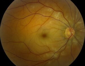
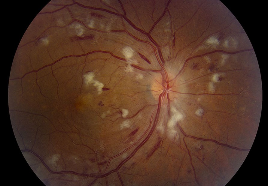
The classic picture includes bilateral retinal hemorrhages, cotton wool spots or disc swelling.
In addition, the brain, kidneys and heart may suffer at the same time.
Why the Eye Shows Damage
A sudden pressure rise can overwhelm autoregulation in retinal and optic nerve vessels.
As a result, endothelial injury and leakage develop.
Choroidal vessels lack comparable autoregulation, so they may become ischemic.
Fundus Explorer Pro
Photograph the retinal findings described here with the phone already in your pocket — 22 D optics and built-in illumination in one handheld unit.
From Choroida — the team behind this siteSymptoms and First Steps
Patients often describe blur, scotomas or headache, usually in both eyes.
However, vision may stay good when the macula is spared.
Measure blood pressure as soon as bilateral hemorrhages or disc swelling appear.
Also ask about chest pain, breathlessness, confusion, weakness and reduced urine output.
Examination Pearls
First, examine both fundi, because findings can be asymmetric.
However, avoid dilation if the team needs to monitor pupils for neurologic change.
Next, look for arteriolar narrowing and arteriovenous crossing changes.
Also note old arteriolar changes, because they suggest long-standing hypertension.
Retinal Signs in Malignant Hypertension
Flame hemorrhages and cotton wool spots reflect nerve fiber layer damage and focal ischemia.
Hard exudates may then form a macular star as leakage persists.
Grading helps description, but symptoms and systemic injury should drive urgency.
Optic Disc Swelling
Bilateral disc edema with severe hypertension is a red flag (see hypertensive optic neuropathy).
The swelling may look identical to papilledema from raised intracranial pressure.
Therefore, check for vomiting, neurologic deficit and altered mental state.
Choroidal Signs in Malignant Hypertension
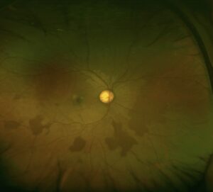
Acute choroidal ischemia can produce pale or yellow deep lesions at the level of the retinal pigment epithelium.
These lesions may later become pigmented Elschnig spots.
In addition, exudative retinal detachment can occur, especially with abrupt pressure rises in younger patients.
OCT and Fluorescein Angiography
OCT can show subretinal fluid, intraretinal fluid and inner retinal ischemic changes.
Fluorescein angiography may reveal delayed choroidal filling and disc leakage.
However, imaging should never delay transfer of an unstable patient.
Same-Day Triage for Malignant Hypertension
Send the patient to the emergency department when severe hypertension accompanies acute fundus changes.
Use ambulance transfer when chest pain, dyspnea, confusion, seizure or focal deficit is present (see same-day eye emergencies).
Then send measured pressures, visual acuity and a description of the fundus with the patient.
Finally, call the receiving physician directly, because a clear handover shortens the delay.
Conditions That Mimic This Picture
Bilateral disc swelling with hemorrhages also occurs with raised intracranial pressure and infiltrative disease.
Similarly, severe anemia, leukemia and radiation retinopathy can cause hemorrhages and cotton wool spots.
Therefore, a normal blood pressure should prompt a broader workup.
Pregnancy and Younger Patients
Preeclampsia can also cause serous retinal detachment and hemorrhages.
Therefore, check blood pressure in any pregnant patient with new visual symptoms.
In addition, secondary causes such as renal disease often explain severe disease in younger adults.
As a result, a normal prior examination does not exclude an acute crisis.
Treatment of Malignant Hypertension
Treatment belongs to the emergency or critical care team, usually with titrated intravenous agents.
For many adults, ACC/AHA guidance advises lowering pressure by no more than 25% within the first hour.
Compelling conditions such as aortic dissection or severe preeclampsia need specific plans.
Visual Risk During Pressure Lowering
Chronic hypertension shifts autoregulation, so abrupt lowering can reduce perfusion to the brain and optic nerve.
Therefore, report any new visual loss during treatment to the treating team immediately.
Antihypertensive therapy treats the cause, but it cannot reverse established optic nerve damage.
Counseling in Malignant Hypertension
Explain that the eye findings signal a systemic emergency, even when vision seems nearly normal.
Fluid and hemorrhages often clear gradually, although recovery may take weeks or months.
Finally, stress medication adherence, because repeated crises can cause further organ damage.
Follow-Up After Malignant Hypertension
Review the patient after stabilization, with timing based on vision and fundus severity.
At each visit, record acuity, color vision, disc appearance and macular OCT.
Also watch for disc pallor, which can indicate lasting optic nerve damage.
Pitfalls to Avoid
- Do not treat bilateral disc swelling as papilledema before checking blood pressure
- Never assume good acuity excludes a hypertensive emergency
- Do not label serous detachment as central serous chorioretinopathy without measuring pressure
- Avoid delaying transfer for angiography


Document what you see
Two smartphone imaging tools built for everyday clinic use — one for the slit lamp, one for the fundus.
From Choroida — the team behind this siteReferences
- Wong TY, Mitchell P. Hypertensive retinopathy. N Engl J Med. 2004;351:2310-2317.
- Wong TY, Mitchell P. The eye in hypertension. Lancet. 2007;369:425-435.
- Hayreh SS, Servais GE, Virdi PS. Fundus lesions in malignant hypertension. V. Hypertensive optic neuropathy. Ophthalmology. 1986;93:74-87.
- Hayreh SS, Servais GE, Virdi PS. Fundus lesions in malignant hypertension. VI. Hypertensive choroidopathy. Ophthalmology. 1986;93:1383-1400.
- Whelton PK, Carey RM, Aronow WS, et al. 2017 ACC/AHA guideline for the prevention, detection, evaluation, and management of high blood pressure in adults. Hypertension. 2018;71:e13-e115.
- van den Born BJH, Lip GYH, Brguljan-Hitij J, et al. ESC Council on hypertension position document on the management of hypertensive emergencies. Eur Heart J Cardiovasc Pharmacother. 2019;5:37-46.

Writes and reviews the clinical articles on Educate. Every post is checked against peer-reviewed sources before it goes live.