Post-LASIK keratitis is a corneal infection, usually centered in the flap interface, after laser in situ keratomileusis.
It is rare, yet it can scar the visual axis quickly.
Therefore, any focal interface infiltrate after LASIK deserves same-day assessment.
What Post-LASIK Keratitis Involves
The flap creates a potential space that can harbor organisms away from the tear film.
As a result, topical drugs may reach the infected site poorly.
In addition, postoperative steroids can blunt inflammation and delay recognition.
How Common Is Post-LASIK Keratitis?
Published series consistently report a low incidence.
For example, Llovet and colleagues found roughly 0.035% in more than 200,000 procedures.
Because most surgeons rarely see a case, late recognition remains a real hazard.
All-fit Slit-Lamp Adapter
Record and share exactly what you see at the slit lamp. One adapter fits any slit lamp or surgical microscope — and any smartphone.
From Choroida — the team behind this siteRisk Factors and Sources
Organisms may come from the lid margin, instruments, or the surgical environment.
Blepharitis and epithelial defects can also add risk.
Likewise, an enhancement that lifts the flap reopens the interface.
Timing of Post-LASIK Keratitis
Time of onset offers the first clue to the likely organism.
Gram-positive bacteria, including staphylococci and streptococci, dominate early disease within the first week or so.
Atypical mycobacteria, fungi, and Nocardia more often cause later, indolent infiltrates.
Therefore, a slowly worsening lesion on a fluoroquinolone should broaden your differential (see fungal and bacterial clues).
Slit Lamp Signs of Post-LASIK Keratitis
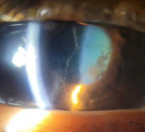
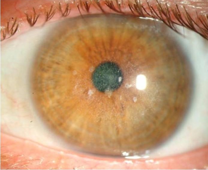
Look first for a focal, creamy, or white infiltrate at the interface.
Then assess conjunctival injection, epithelial defects, flap edema, and anterior chamber reaction.
Satellite lesions suggest a fungal cause, while a crystalline or “cracked windshield” look can suggest mycobacteria.
However, symptoms may seem mild on steroids, and reduced corneal sensation can dampen pain.
Finally, document size and depth, because small changes guide the next decision.
Post-LASIK Keratitis Versus DLK
Diffuse lamellar keratitis is a sterile inflammatory reaction confined to the interface.
It usually appears within the first few days as fine, diffuse, granular haze with minimal pain.
Infection, however, tends to be focal, dense, and progressive, with more pain and injection.
Infection may also look quieter on steroids, so early improvement does not exclude it.
Other Mimics to Exclude
Pressure-induced stromal keratitis can resemble DLK, usually on steroids, with elevated intraocular pressure.
Epithelial ingrowth typically forms pearly nests or sheets near the flap edge (see flap complications).
Herpes simplex reactivation can also occur after surgery.
Thus, check pressure, inspect the flap edge, and review any herpetic history.
Workup for Post-LASIK Keratitis
Significant interface infiltrates usually call for lifting the flap in a clean setting.
Next, scrape the interface and send material for smears and cultures.
Request fungal media and mycobacterial studies, particularly in delayed disease.
Also consider confocal microscopy or anterior segment OCT to define depth.
Treating Post-LASIK Keratitis
After sampling, many specialists irrigate the interface with antibiotic solution to reduce organism load.
Frequent topical therapy then follows, based on onset and smear findings.
The ASCRS white paper favors broad gram-positive coverage for early disease, with amikacin-based coverage for delayed disease.
Steroids usually stop while infection remains active.
Even so, antibiotics cannot reverse established scarring or irregular astigmatism.
When the Flap Must Go
Persistent infection may require flap amputation, which removes infected tissue and improves drug penetration.
However, amputation can induce haze and a significant refractive shift.
In addition, progressive melting or perforation may require keratoplasty.
Follow-Up After Post-LASIK Keratitis
First, examine the patient daily until the infiltrate clearly shrinks.
Then space visits as the epithelium heals and the interface clears.
Also record visual acuity and measure the infiltrate at every visit.
However, a stable size does not always mean control, so watch for new satellite lesions.
Finally, taper steroids only after cultures and clinical response support it.
Prevention of Post-LASIK Keratitis
Treat blepharitis before surgery, because lid flora can seed the interface.
In addition, maintain strict sterile technique and clean all instruments carefully.
Next, advise patients to avoid water exposure and eye rubbing in the first weeks.
Therefore, give clear written instructions about pain, redness, and falling vision.
Also stress that these symptoms need a same-day call.
Counseling in Post-LASIK Keratitis
Explain that infection is uncommon but can threaten vision without prompt treatment.
Next, warn patients that frequent drops and repeated visits may continue for weeks.
Final vision depends on organism, location, and scarring, so avoid early promises.
Pitfalls to Avoid
- Do not label every interface haze as DLK without checking for focal infiltrates
- Never escalate steroids without a clear plan for close reexamination
- Do not start empirical treatment before sampling a significant lesion, when sampling is feasible
- Avoid dismissing late symptoms, because atypical organisms can appear months after surgery


Document what you see
Two smartphone imaging tools built for everyday clinic use — one for the slit lamp, one for the fundus.
From Choroida — the team behind this siteReferences
- Chang MA, Jain S, Azar DT. Infections following laser in situ keratomileusis: an integration of the published literature. Surv Ophthalmol. 2004;49:269-280.
- Karp CL, Tuli SS, Yoo SH, et al. Infectious keratitis after LASIK. Ophthalmology. 2003;110:503-510.
- Llovet F, de Rojas V, Interlandi E, et al. Infectious keratitis in 204 586 LASIK procedures. Ophthalmology. 2010;117:232-238.
- Donnenfeld ED, Kim T, Holland EJ, et al. ASCRS White Paper: management of infectious keratitis following laser in situ keratomileusis. J Cataract Refract Surg. 2005;31:2008-2011.
- Smith RJ, Maloney RK. Diffuse lamellar keratitis: a new syndrome in lamellar refractive surgery. Ophthalmology. 1998;105:1721-1726.

Writes and reviews the clinical articles on Educate. Every post is checked against peer-reviewed sources before it goes live.