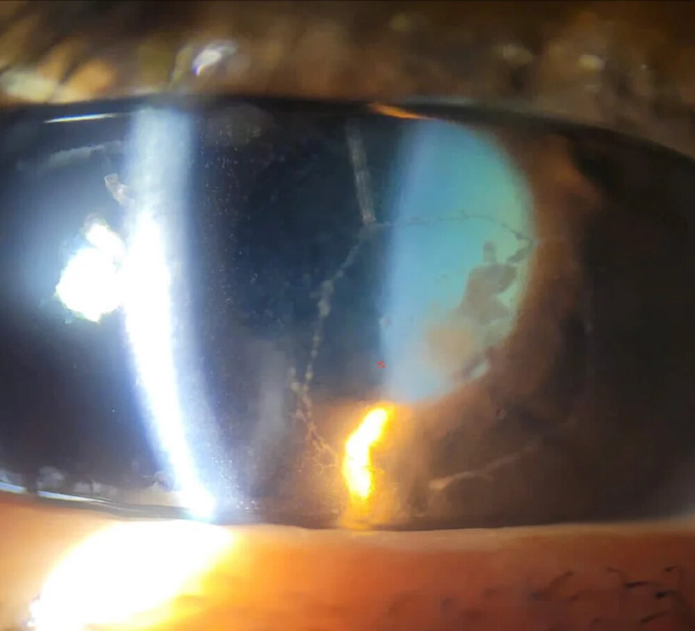
Endothelial plaque is a white or gray exudate adherent to the corneal endothelium, classically beneath a fungal ulcer.
It suggests that disease has reached the deep stroma, Descemet membrane, or anterior chamber.
However, the sign is suggestive and does not prove fungal infection.
What Endothelial Plaque Looks Like
On slit lamp examination, the plaque appears as a thick, immobile deposit on the posterior cornea.
It usually lies directly behind the stromal infiltrate.
Its surface also often looks fluffy or fibrinous, while the margins may be irregular.
A narrow slit beam then separates the plaque from the overlying stroma.
Why the Plaque Forms
Filamentous fungi can extend through the stroma and may cross Descemet membrane.
As a result, fibrin, inflammatory cells, and sometimes organisms collect on the posterior surface.
Therefore, a plaque may represent infection, inflammation, or both.
All-fit Slit-Lamp Adapter
Record and share exactly what you see at the slit lamp. One adapter fits any slit lamp or surgical microscope — and any smartphone.
From Choroida — the team behind this siteEndothelial Plaque and Depth
A plaque under a superficial-looking ulcer should prompt reassessment of depth.
For example, the epithelial defect may be small while deep infection continues beneath it.
Next, check for stromal thinning, descemetocele, and anterior chamber involvement.
Examination Pearls
First, use a narrow slit beam at an oblique angle to separate the plaque from stromal edema.
Next, grade anterior chamber cell, flare, and fibrin, because they guide the severity assessment.
Also check intraocular pressure, since inflammation can change it in either direction.
Then photograph the plaque at baseline so that follow-up comparison is objective.
Associated Findings
Patients usually report pain, redness, photophobia, and reduced vision.
In addition, a thick, immobile hypopyon often accompanies the plaque.
Feathery margins, a dry raised surface, and satellite lesions increase suspicion of a filamentous fungus.
However, no single sign establishes the organism (see fungal versus bacterial clues).
Imaging an Endothelial Plaque
Anterior segment OCT can show a hyperreflective mass on the posterior corneal surface.
It also helps estimate residual stromal thickness when the slit lamp view is poor.
Finally, confocal microscopy may reveal filaments within the stroma, although it depends on operator skill.
Differential Diagnosis of Endothelial Plaque
Several conditions can produce material on the posterior cornea:
- Severe bacterial keratitis with deep extension
- Herpetic endotheliitis with keratic precipitates
- Graft rejection with an endothelial rejection line
- Endophthalmitis with fibrin on the endothelium
- Retained lens material after cataract surgery
Confirming the Diagnosis
First, send corneal scrapings for smear and fungal culture before changing therapy when possible.
However, surface samples can be negative when organisms lie deep in the stroma.
A corneal biopsy may then improve the yield.
Also review prior antibiotic or steroid use because it can alter the picture.
Endothelial Plaque and Topical Therapy
Natamycin 5% is a standard first choice for filamentous keratitis.
In MUTT I, natamycin produced better outcomes than topical voriconazole, especially in Fusarium cases.
However, natamycin penetrates deep tissue poorly.
A plaque therefore raises concern that topical drug may not reach all of the infection.
Epithelial debridement may improve drug entry, although it cannot clear endothelial disease.
Systemic and Targeted Therapy
MUTT II found no overall benefit from adding oral voriconazole in severe filamentous ulcers.
A subgroup analysis suggested possible benefit in Fusarium ulcers, but that needs cautious interpretation.
Intrastromal or intracameral antifungals deliver drug closer to the lesion.
However, the evidence comes from small studies, and Sharma and colleagues found no advantage of intrastromal voriconazole as an adjunct.
Therefore, discuss injections as an option with uncertain benefit.
Steroids and Endothelial Plaque
Avoid corticosteroids while active fungal infection is suspected.
Steroids can reduce visible inflammation while fungal growth continues.
As a result, a smaller plaque on steroids does not confirm microbial control.
Risk of Endophthalmitis
Fungal extension into the anterior chamber can progress to endophthalmitis.
Therefore, ask about floaters and examine the vitreous whenever the view allows.
If the view is poor, B-scan ultrasonography can then detect vitreous opacities.
In addition, vitreous involvement changes management and requires urgent retina consultation.
When Surgery Is Needed
A plaque alone does not mandate keratoplasty.
However, enlarging infiltrates, progressive thinning, perforation, or limbal spread strengthen the case for therapeutic keratoplasty.
Graft failure, recurrence, cataract, and glaucoma remain important risks.
Monitoring and Counseling
Measure the infiltrate, epithelial defect, hypopyon, and plaque at each visit.
A plaque may resolve slowly, even during effective therapy.
Therefore, interpret it alongside pain, stromal density, and thinning.
Explain that treatment may take weeks and that frequent drops are necessary.
Also warn about perforation, scarring, and possible transplantation, and ask patients to return urgently if pain worsens.
Pitfalls to Avoid
- Do not assume every endothelial plaque is fungal
- Do not judge depth from the epithelial defect alone
- Avoid stopping antifungals because the surface looks better
- Never start steroids before excluding fungal disease


Document what you see
Two smartphone imaging tools built for everyday clinic use — one for the slit lamp, one for the fundus.
From Choroida — the team behind this siteReferences
- Thomas PA, Leck AK, Myatt M. Characteristic clinical features as an aid to the diagnosis of suppurative keratitis. Br J Ophthalmol. 2005;89:1554-1558.
- Prajna NV, Krishnan T, Mascarenhas J, et al. The Mycotic Ulcer Treatment Trial: a randomized trial comparing natamycin vs voriconazole. JAMA Ophthalmol. 2013;131:422-429.
- Prajna NV, Krishnan T, Rajaraman R, et al. Effect of oral voriconazole on fungal keratitis in the Mycotic Ulcer Treatment Trial II (MUTT II). JAMA Ophthalmol. 2016;134:1365-1372.
- Sharma N, Chacko J, Velpandian T, et al. Comparative evaluation of topical versus intrastromal voriconazole as an adjunct to natamycin in recalcitrant fungal keratitis. Ophthalmology. 2013;120:677-681.
- Thomas PA, Kaliamurthy J. Mycotic keratitis: epidemiology, diagnosis and management. Clin Microbiol Infect. 2013;19:210-220.

Writes and reviews the clinical articles on Educate. Every post is checked against peer-reviewed sources before it goes live.