
Nontuberculous mycobacterial keratitis (NTM keratitis) is a slowly progressive corneal infection caused by environmental mycobacteria, most often Mycobacterium chelonae and Mycobacterium abscessus.
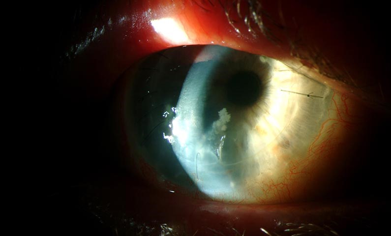
It follows refractive surgery, trauma with soil or water contamination, and contact lens wear, and it is frequently misdiagnosed as a fungal or herpetic infection.
The delay in diagnosis is the main problem, and the key step is to think of the organism.
What Is Nontuberculous Mycobacterial Keratitis?
These organisms live in water, soil, and dust, and they are resistant to many disinfectants and antibiotics.
Infection usually occurs through a break in the epithelium or through a surgical wound, and the course is measured in weeks and not in days.
Post-LASIK infections have been reported in clusters, which points to a contaminated source such as water or instruments.
The infection is uncommon in the general population, but it has a clear tendency to follow procedures in which the stromal interface is exposed.
Clinical Features
The patient has pain and redness that are often out of proportion to the early findings, or sometimes less than expected.
The infiltrate is gray-white, often with a ground-glass appearance and irregular, feathery or wreath-like margins.
Satellite lesions may be present, which can resemble fungal keratitis.
In LASIK cases the infiltrate lies at the flap interface, and it can spread laterally beneath the flap.
Anterior chamber reaction is usually mild, and hypopyon is uncommon until late in the disease.
Corneal melting occurs if the infection is not controlled.
All-fit Slit-Lamp Adapter
Record and share exactly what you see at the slit lamp. One adapter fits any slit lamp or surgical microscope — and any smartphone.
From Choroida — the team behind this siteDiagnosis
A scraping for smear and culture is needed, and the laboratory should be told that mycobacteria are suspected.
Acid-fast stains, such as Ziehl-Neelsen or auramine-rhodamine, can show the organisms, and cultures on Lowenstein-Jensen or Middlebrook media should be requested.
Growth takes days to weeks, so treatment is often started before the result is available.
Confocal microscopy can help to show organisms in the stroma, and PCR is useful when it is available.
When the interface is involved, lifting the flap to obtain a sample is sometimes needed.
Differential Diagnosis
- Fungal keratitis: feathery edges and satellites, usually with a history of plant trauma (see fungal versus bacterial keratitis)
- Herpetic stromal keratitis: reduced sensation, dendritic history, and a response to antivirals
- Acanthamoeba keratitis: severe pain and a ring infiltrate (see Acanthamoeba keratitis)
- Diffuse lamellar keratitis: sterile, early after LASIK, and responsive to steroids
Treatment
Amikacin is the traditional first-line agent, used as a topical fortified preparation, often together with a macrolide such as clarithromycin or azithromycin.
Fluoroquinolones such as moxifloxacin have activity against some species, and the choice should be guided by susceptibility testing.
Treatment is prolonged, often for months, and a flap lift with irrigation or flap amputation may be needed for interface disease.
Topical steroids are avoided until the infection is controlled.
Deep infections that do not respond may need therapeutic keratoplasty.
Patients must be told that the course is long and that follow-up should be close.
Why the Diagnosis Is Often Late
The infiltrate evolves slowly, and in the first weeks it may look like a mild, culture-negative infection that improves briefly on a fluoroquinolone and then worsens.
The early response to a broad-spectrum antibiotic can mislead the clinician, because some fluoroquinolones have partial activity against the organism.
Several clinicians have noted that the interface infiltrate after LASIK may be misread as diffuse lamellar keratitis or as a sterile reaction, and that the use of steroids in that setting makes the infection worse.
A high index of suspicion is therefore needed in any post-LASIK patient who has a late infiltrate, particularly when other cases have been seen from the same surgical center.
Risk Factors and Prevention
Exposure to contaminated water, instruments, or solutions is the main link in postoperative clusters.
Contact lens wearers who shower or swim in lenses are at risk, and so are patients with a history of trauma in a contaminated environment.
Surgical centers should follow strict sterilization routines and should report clusters of delayed postoperative infections to the public health authority.
Patients should be advised to avoid water exposure in the early postoperative period and to report increasing pain or redness promptly.
Prognosis
With early diagnosis and a prolonged course of appropriate antibiotics, many eyes heal with a scar and acceptable vision.
Delayed treatment leads to stromal necrosis, and some eyes need keratoplasty.
Follow-up should continue for several months after the infiltrate has cleared, because relapse after stopping treatment has been described.


Document what you see
Two smartphone imaging tools built for everyday clinic use — one for the slit lamp, one for the fundus.
From Choroida — the team behind this siteReferences
- Chandra NS, Torres MF, Winthrop KL, et al. Cluster of Mycobacterium chelonae keratitis cases following laser in-situ keratomileusis. Am J Ophthalmol. 2001;132:819-830.
- Girgis DO, Karp CL, Miller D. Ocular infections caused by non-tuberculous mycobacteria: update on epidemiology and management. Clin Exp Ophthalmol. 2012;40:467-475.
- Mannis MJ, Holland EJ, eds. Cornea. 4th ed. Elsevier; 2017.
- American Academy of Ophthalmology. Basic and Clinical Science Course, Section 8: External Disease and Cornea. AAO; current edition.

Writes and reviews the clinical articles on Educate. Every post is checked against peer-reviewed sources before it goes live.


