
Introduction
Microbial keratitis is one of the most vision-threatening ophthalmic emergencies encountered in daily practice.

Early diagnosis and prompt treatment are essential to prevent corneal scarring, perforation, and permanent visual loss.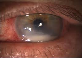
However, differentiating between fungal keratitis and bacterial keratitis can sometimes be clinically challenging, especially during the early stages of disease.
Mistaking fungal keratitis for bacterial keratitis is a common and dangerous pitfall. Delayed antifungal therapy often leads to progressive stromal destruction and poor visual outcomes.
Conversely, overdiagnosing fungal infection may expose patients to prolonged, potentially toxic treatment unnecessarily.
For ophthalmologists, recognizing the subtle but critical clinical differences between these two entities can significantly alter patient prognosis.
Why Differentiation Matters
The treatment strategies for fungal and bacterial keratitis are fundamentally different.
Bacterial Keratitis:
- Usually progresses rapidly
- Often responds well to intensive topical antibiotics
- Steroids may later play a role after infection control
Fungal Keratitis:
- Typically more indolent
- Requires prolonged antifungal therapy
- Often worsens with inappropriate steroid use
- Associated with a higher risk of perforation
👉 Delayed diagnosis of fungal keratitis remains one of the leading causes of treatment failure in corneal infections.
Epidemiology and Risk Factors
Understanding patient history is often the first diagnostic clue.
Common Risk Factors for Fungal Keratitis:
- Trauma with vegetative matter
- Agricultural or outdoor injuries
- Chronic ocular surface disease
- Topical steroid abuse
- Warm, humid climates
- Contact lens wear (less common but increasing)
Common Risk Factors for Bacterial Keratitis:
- Contact lens misuse
- Ocular surface disease
- Previous ocular surgery
- Corneal epithelial defects
- Chronic blepharitis
👉 A history of trauma with plant material should immediately raise suspicion for fungal infection.
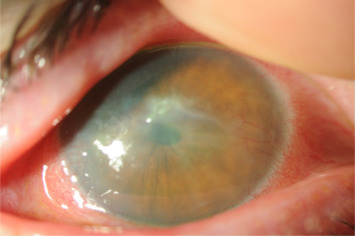
Clinical Appearance: The Most Important Clues
1. Infiltrate Margins
Fungal Keratitis:
Typically presents with:
- Feathery or irregular borders
- Dry, gray-white stromal infiltrate
- Satellite lesions surrounding the main ulcer
Bacterial Keratitis:
Usually demonstrates:
- Dense suppurative infiltrate
- Well-defined borders
- More localized appearance
👉 Satellite lesions are highly suggestive of fungal infection.
2. Surface Texture
Fungal Keratitis:
- Dry or rough corneal surface
- Elevated slough or plaque-like appearance
Bacterial Keratitis:
- Wet, purulent appearance
- Dense stromal suppuration
The “dry-looking ulcer” is a classic fungal clue many clinicians remember.
3. Progression Speed
Bacterial Keratitis:
- Rapid onset and progression
- Severe pain and redness for a short duration
Fungal Keratitis:
- Slower progression
- Symptoms may appear deceptively mild early on
However, fungal infections can become extremely aggressive once deep stromal invasion occurs.
4. Hypopyon Characteristics
Both fungal and bacterial keratitis may present with hypopyon, but there are subtle differences.
Fungal Hypopyon:
- Often immobile or fixed
- Thick and fibrinous
Bacterial Hypopyon:
- More fluid and mobile
While not diagnostic alone, this finding can support clinical suspicion.
5. Pain Severity
Bacterial Keratitis:
Pain is often severe and disproportionate to lesion size.
Fungal Keratitis:
Pain may initially be surprisingly mild despite significant stromal involvement.
This discrepancy sometimes delays patient presentation and diagnosis.

Special Organisms and Unique Presentations
Filamentous Fungi
Common organisms include:
- Fusarium
- Aspergillus
Typical features:
- Feathery infiltrates
- Satellite lesions
- Endothelial plaque
Yeast Infections (Candida)
More common in:
- Chronically diseased corneas
- Immunocompromised patients
May resemble bacterial keratitis more closely, making diagnosis difficult.
Role of Corneal Scraping and Microbiology
Clinical examination alone is sometimes insufficient.
Essential Diagnostic Tools:
- Gram stain
- KOH wet mount
- Culture media
- Confocal microscopy (when available)
Why Early Scraping Matters:
- Identifies atypical organisms
- Prevents treatment delay
- Guides targeted therapy
👉 Any non-responding keratitis should undergo repeat microbiological evaluation.
Steroids: A Critical Warning
One of the most dangerous mistakes in ophthalmology is the inappropriate use of steroids in undiagnosed fungal keratitis.
Steroids Can:
- Suppress local immunity
- Accelerate fungal proliferation
- Mask clinical progression
- Increase perforation risk
While steroids may later benefit selected bacterial keratitis cases, they should be used with extreme caution in fungal disease.
Imaging and Advanced Diagnostic Clues
Anterior segment OCT and confocal microscopy are increasingly valuable.
Confocal Microscopy May Show:
- Branching fungal filaments
- Deep stromal invasion
AS-OCT Can Demonstrate:
- Stromal necrosis
- Depth of infiltrate
- Endothelial plaques
These technologies are especially useful in equivocal or deep infections.
Treatment Differences
Bacterial Keratitis:
Mainstay treatment includes:
- Fortified topical antibiotics
- Fluoroquinolones
- Cycloplegics
- Adjunctive steroids in selected cases
Fungal Keratitis:
Treatment often requires:
- Topical natamycin
- Voriconazole
- Systemic antifungals in severe cases
- Therapeutic keratoplasty in advanced disease
Fungal ulcers generally require a much longer treatment duration compared to bacterial ulcers.
When to Suspect Fungal Keratitis Immediately
Ophthalmologists should maintain high suspicion when encountering:
- Feathery infiltrates
- Satellite lesions
- History of vegetative trauma
- Dry elevated ulcer
- Poor response to antibiotics
- Fixed hypopyon
- Chronic indolent progression
Recognizing these clues early can be vision-saving.
Conclusion
Differentiating fungal from bacterial keratitis is one of the most important clinical skills in corneal disease management. Although overlap exists, careful attention to patient history, infiltrate characteristics, progression pattern, and microbiological testing can greatly improve diagnostic accuracy.
For ophthalmologists, the key is not simply identifying infection—but identifying the right infection before irreversible damage occurs.
Early suspicion, timely scraping, and appropriate therapy remain the cornerstone of successful management.
References
- Thomas PA, Kaliamurthy J. “Mycotic keratitis: epidemiology, diagnosis and management.” Clin Microbiol Infect. 2013.
- Ting DSJ, et al. “Infectious keratitis: an update on epidemiology and management.” Eye (Lond). 2021.
- Alfonso EC, et al. “Fungal keratitis.” Lancet. 2019.
- Sharma N, et al. “Evaluation of clinical and microbiological profile of microbial keratitis.” Br J Ophthalmol. 2010.
- Wilhelmus KR. “Indecision about corticosteroids for bacterial keratitis.” Ophthalmology. 2002.
- Srinivasan M. “Fungal keratitis.” Curr Opin Ophthalmol. 2004.
- Dart JK, et al. “Microbial keratitis: current management strategies.” Eye. 2015.
- Garg P. “Diagnosis of microbial keratitis.” Br J Ophthalmol. 2010.
