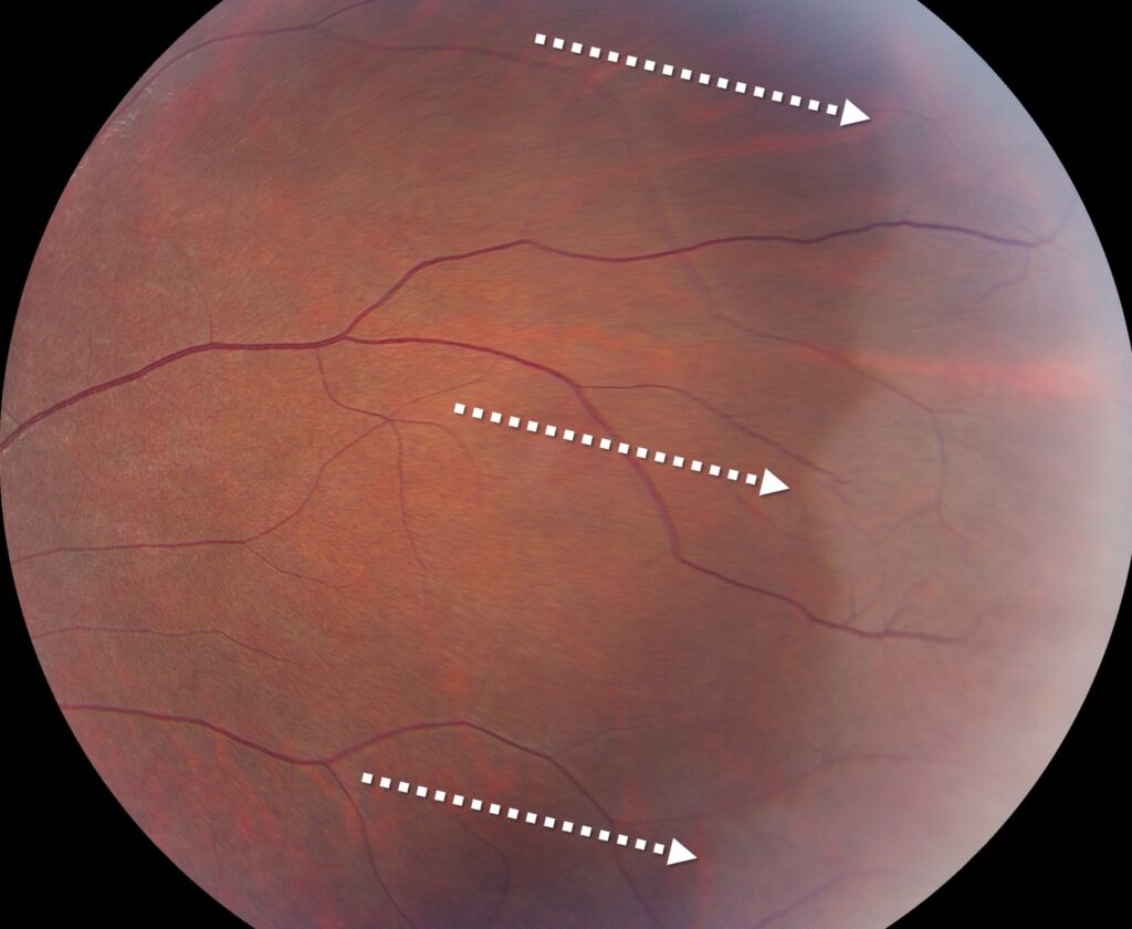
White with pressure is a translucent, gray-white change in the peripheral retina that appears during scleral depression.
Examiners usually find it incidentally during a dilated peripheral examination.
Because white without pressure looks similar, clinicians often confuse the two signs.
What White With Pressure Means
The term describes a change in retinal color under indentation.
When the depressor raises the eye wall, the affected retina loses some of its normal transparency.
Once the depressor moves away, the area returns to its usual appearance.
Therefore, the sign depends on the examiner’s maneuver.
How White With Pressure Looks
The whitening usually forms a patch or band in the far periphery.
Its posterior edge may look distinct, while its anterior part often blends toward the ora serrata.
It may also span several clock hours.
In addition, the retina stays attached, and the overlying vessels keep their normal course.
Fundus Explorer Pro
Photograph the retinal findings described here with the phone already in your pocket — 22 D optics and built-in illumination in one handheld unit.
From Choroida — the team behind this siteWhy the Sign Appears
Watzke published a clinicopathologic correlation of this sign in 1961.
Later discussions have linked peripheral whitening with the vitreoretinal interface and vitreous traction.
However, the exact mechanism remains uncertain.
Therefore, treat the name as a clinical description, without assuming a single pathology.
Who Shows White With Pressure
Healthy eyes commonly show this sign, including eyes of young patients.
Karlin and Curtin also related peripheral whitening to greater axial length in myopic eyes.
Pigmented fundi may make the contrast easier to see.
Consequently, its presence alone says little about individual risk.
White With Pressure vs White Without Pressure
The main clinical difference lies in the need for indentation.
White with pressure requires depression, while white without pressure remains visible during ordinary indirect ophthalmoscopy.
Some authors view them as related points on one spectrum.
Also, one eye may show both signs in neighboring areas.
Neither finding alone calls for treatment.
Finding the Sign on Examination
First, dilate the pupil fully and examine each quadrant systematically.
Next, use gentle, controlled indentation and watch the retina change as the depressor moves (see scleral depression technique).
Dynamic viewing also helps because a flat whitening shifts differently from a true break.
Finally, compare the fellow eye, since these changes are often bilateral.
Symptoms and White With Pressure
Patients do not report symptoms from this sign.
Therefore, new flashes, floaters or a field shadow need another explanation.
Acute posterior vitreous detachment, retinal tear and detachment must then be excluded.
A careful examination should answer that question before you offer reassurance.
Differential Diagnosis
Several peripheral findings can resemble this whitening.
- White without pressure, visible with no indentation
- Lattice degeneration, often with sclerotic vessels or pigment
- Retinal tear, with a visible defect or flap
- Degenerative retinoschisis, with a smooth, elevated inner layer
- Shallow retinal detachment, with subretinal fluid
- Commotio retinae, after recent blunt trauma
Benign variants can also add confusion, such as meridional folds and cobblestone degeneration.
When White With Pressure Needs Closer Review
The sign itself rarely drives the plan, but its clinical setting can.
Examine the periphery more carefully in these situations:
- New flashes, floaters or a field defect
- High myopia or long axial length
- Previous tear or detachment in either eye
- Family history of retinal detachment
- Recent ocular trauma
Retinal Breaks and Peripheral Whitening
Some clinicians have described breaks near the posterior margin of peripheral whitening.
Freeman also studied fellow eyes of patients with giant retinal breaks, where peripheral changes can draw attention.
However, this association does not prove that the sign causes tears.
Therefore, inspect the posterior border closely, especially in a high-risk eye.
Managing White With Pressure
Isolated white with pressure needs no treatment.
Prophylactic laser has no established role for this sign alone, and it cannot eliminate future detachment risk.
Laser also carries its own risks, so it needs a clear indication.
If you find a break, then manage that lesion according to its type, symptoms and risk features.
Follow-Up Planning
No evidence supports a fixed schedule for this isolated finding.
Therefore, routine eye care usually suffices for an asymptomatic patient.
However, high-risk eyes may need a shorter interval based on the overall picture.
Repeat depression whenever symptoms change, because an earlier normal examination cannot exclude a new tear.
Counseling Patients
Explain that the examination revealed a common peripheral finding, and that it does not need treatment.
Next, describe the warning symptoms in plain language.
Ask the patient to seek same-day care for new floaters, flashes, a curtain or sudden blur.
This approach gives reassurance while keeping the safety message clear.
Pitfalls to Avoid
- Do not call the sign a retinal tear without seeing a defect
- Never let this label explain new symptoms
- Avoid laser for the sign alone
- Do not skip the posterior border
- Avoid heavy indentation that makes interpretation harder
For the latest studies, see the PubMed results for white with pressure.


Document what you see
Two smartphone imaging tools built for everyday clinic use — one for the slit lamp, one for the fundus.
From Choroida — the team behind this siteReferences
- Watzke RC. The ophthalmoscopic sign “white with pressure”: a clinicopathologic correlation. Arch Ophthalmol. 1961;66:812-823.
- Karlin DB, Curtin BJ. Peripheral chorioretinal lesions and axial length of the myopic eye. Am J Ophthalmol. 1976;81:625-635.
- Freeman HM. Fellow eyes of giant retinal breaks. Trans Am Ophthalmol Soc. 1978;76:343-382.
- Orlin A, Fatoo A, Ehrlich J, et al. Ultra-widefield fluorescein angiography of white without pressure. Clin Ophthalmol. 2013;7:959-964.
- Flaxel CJ, Adelman RA, Bailey ST, et al. Posterior vitreous detachment, retinal breaks, and lattice degeneration Preferred Practice Pattern. Ophthalmology. 2020;127:P146-P181.

Writes and reviews the clinical articles on Educate. Every post is checked against peer-reviewed sources before it goes live.