
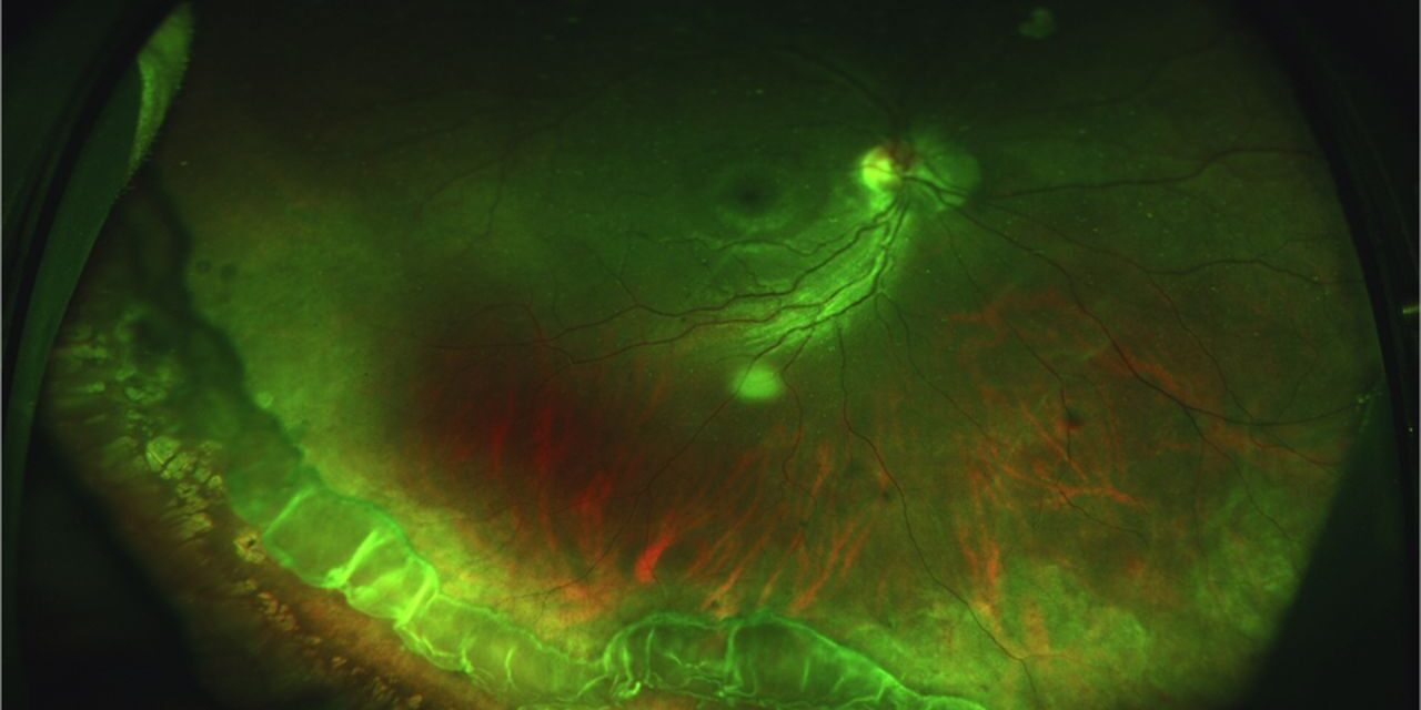
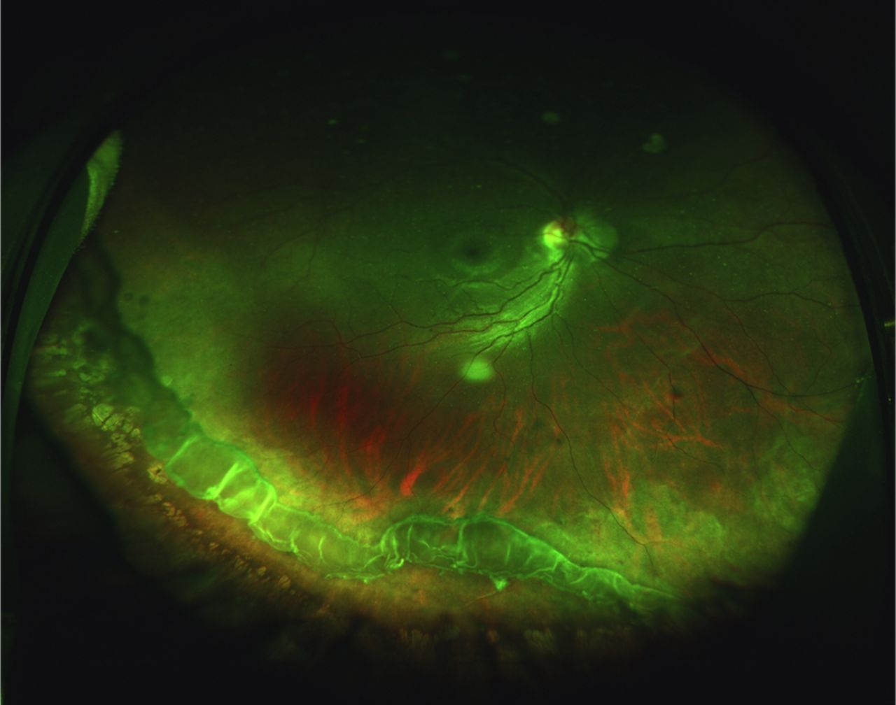
Vitreous base avulsion is a specific, traumatic finding at the very periphery of the retina, and recognizing its distinctive appearance, a torn, often mobile strand of tissue avulsed directly from the ora serrata region, matters because this finding carries a genuinely high risk of associated retinal detachment, making its correct identification during trauma evaluation a priority rather than an incidental note.
What Gets Torn
The vitreous base is a firm, circumferential zone of especially tight vitreoretinal adhesion spanning the ora serrata and the immediately adjacent retina and pars plana, and significant blunt trauma to the eye can generate enough shearing and traction force at this zone of unusually strong adhesion to tear tissue away entirely, avulsing a strip of the vitreous base, often including adjacent non-pigmented pars plana epithelium and peripheral retina, from its normal position.
The Bucket-Handle Appearance
Once avulsed, the torn strip of tissue, still attached to the vitreous, often remains visible as a mobile, elevated band or strand within the peripheral vitreous cavity, classically described as resembling the handle of a bucket given its arched, strand-like appearance connecting two points along the peripheral retina, and this distinctive shape is one of the more specific and recognizable signs of significant peripheral traumatic vitreoretinal injury.
Fundus Explorer Pro
Photograph the retinal findings described here with the phone already in your pocket — 22 D optics and built-in illumination in one handheld unit.
From Choroida — the team behind this siteAssociated Findings
- A retinal dialysis, a specific type of peripheral retinal break in which the retina separates cleanly at the ora serrata, is frequently associated with vitreous base avulsion, since both injuries result from the same underlying traumatic shearing mechanism at this zone of firm adhesion
- Vitreous hemorrhage is common given the traumatic mechanism, and can sometimes limit the view of the peripheral retina, requiring careful examination once any hemorrhage has cleared sufficiently, or B-scan ultrasound in the interim
- Other signs of significant blunt trauma, including angle recession, iris sphincter tears, or lens subluxation, may be present concurrently, reflecting the same underlying traumatic event (see angle recession glaucoma)
Clinical Significance and Detachment Risk
Vitreous base avulsion, particularly when associated with a retinal dialysis, carries a significant risk of subsequent retinal detachment, and this risk can be somewhat delayed relative to the original traumatic event, since a dialysis can progress gradually over weeks to months before producing symptomatic, clinically apparent detachment, in contrast to the more immediately symptomatic presentation typical of an acute flap tear from posterior vitreous detachment.
This delayed, sometimes insidious progression is precisely why any patient with known significant blunt ocular trauma warrants a thorough peripheral retinal examination, with scleral depression, at the time of injury, rather than only if or when symptoms of detachment eventually develop.
Diagnostic Evaluation
- A history of significant blunt trauma to the eye or orbit is the essential context prompting specific attention to this finding
- Dilated fundus examination with careful scleral depression to adequately visualize the far peripheral retina and ora serrata region where these findings occur
- When vitreous hemorrhage limits the direct view, B-scan ultrasound can sometimes identify the mobile, avulsed tissue strand and assess for any associated retinal detachment, supporting decision-making about the timing and urgency of further examination once media clarity improves
- Examination of the fellow eye is also appropriate given that significant trauma, particularly from a blast or diffuse mechanism, can sometimes affect both eyes
Management
Associated Retinal Break
When a retinal dialysis or other break is identified in association with vitreous base avulsion, prophylactic laser retinopexy or cryotherapy is applied around the break, following standard principles for treating any retinal break with meaningful detachment risk, aiming to create a chorioretinal adhesion before subretinal fluid can accumulate and spread.
Vitreous Base Avulsion Without an Identified Break
Even when the avulsed tissue itself is identified without any clearly associated break, close, sustained follow-up is warranted given the recognized risk of a dialysis developing or being identified on subsequent, repeated careful examination, since an initial examination can sometimes miss a subtle break, particularly with any residual vitreous hemorrhage limiting full visualization.
Managing Vitreous Hemorrhage
Vitreous hemorrhage associated with the traumatic injury is generally managed conservatively initially, allowing time for clearing, with surgical vitrectomy considered if hemorrhage fails to clear adequately or if it is preventing necessary examination and treatment of an associated retinal break.
Prognosis
With prompt recognition and appropriate treatment of any associated retinal break, the risk of progression to clinically significant retinal detachment is substantially reduced, underscoring the value of a complete, careful peripheral retinal examination as a standard part of evaluating any patient with significant blunt ocular trauma.
Delayed presentation, when a dialysis has progressed to established retinal detachment before being identified, generally requires more extensive surgical repair and carries a correspondingly more guarded visual prognosis than detection and treatment at the earlier, pre-detachment stage.


Document what you see
Two smartphone imaging tools built for everyday clinic use — one for the slit lamp, one for the fundus.
From Choroida — the team behind this siteReferences
- Cox MS, Schepens CL, Freeman HM. Retinal detachment due to ocular contusion. Arch Ophthalmol. 1966;76:678-685.
- Goffstein R, Burton TC. Differentiating traumatic from nontraumatic retinal detachment. Ophthalmology. 1982;89:361-368.
- Ross WH. Traumatic retinal dialyses. Arch Ophthalmol. 1981;99:1371-1374.
- Johnston PB. Traumatic retinal detachment. Br J Ophthalmol. 1991;75:18-21.
