Corneal scraping collects material from an infectious corneal ulcer for stains and cultures.
It identifies the organism and guides therapy when empirical treatment fails.
Therefore, learn to do it cleanly, since the sample is only as good as the technique.
When to Perform Corneal Scraping
Scrape large or central ulcers, ulcers with a hypopyon, and chronic or atypical infiltrates.
Also, scrape ulcers that do not respond to empirical therapy.
Small peripheral infiltrates can often be treated empirically without a sample.
Because Preferred Practice Patterns support this approach, use clinical judgment.
Preparing the Patient and the Tray
Explain the procedure and use topical anesthesia.
Some anesthetics contain preservatives that can reduce growth, so use a small amount.
Prepare a sterile blade or a Kimura spatula, slides, and culture plates before you begin.
Also, wear gloves and work under the slit lamp.
All-fit Slit-Lamp Adapter
Record and share exactly what you see at the slit lamp. One adapter fits any slit lamp or surgical microscope — and any smartphone.
From Choroida — the team behind this siteTechnique of Corneal Scraping
Scrape the leading edge and the base of the ulcer with firm, gentle strokes.
Use a new, sterile instrument for each medium if possible.
For suspected fungal disease, sample deeper layers.
Because the cornea may be thin, stay gentle and avoid perforation.
Media and Stains
Plate the material on blood agar, chocolate agar and Sabouraud agar.
Use thioglycollate broth to enrich the sample.
For Acanthamoeba, use non-nutrient agar with an Escherichia coli overlay (see Acanthamoeba keratitis).
Also, prepare slides for Gram, Giemsa and calcofluor white or potassium hydroxide stains.
Interpreting Corneal Scraping Results
A Gram stain gives a rapid clue within the hour.
Cultures take days, and fungal growth may need a week or longer.
Also, culture yield varies and a negative culture does not exclude infection.
Therefore, correlate the result with the clinical picture (see fungal versus bacterial keratitis).
Contact Lenses and Solutions
In contact lens wearers, also culture the lens, the case and the solution.
These samples may identify Pseudomonas or Acanthamoeba (see Pseudomonas keratitis).
Because the case often carries the organism, ask the patient to bring it.
Safety of Corneal Scraping
Perforation is rare, but it can occur in a very thin cornea.
Also, avoid scraping a descemetocele.
Use disposable instruments, and dispose of sharps safely.
Because infection spreads through contaminated hands, clean the area afterwards.
After the Sample
Start empirical therapy immediately and adjust it when the results arrive.
Review the patient within 24 hours.
Also, polymerase chain reaction and confocal microscopy help when cultures fail.
Fungal Keratitis and Deeper Sampling
Fungal ulcers extend into the stroma, so a superficial scrape may miss the organism.
Also, a deeper scrape or a corneal biopsy helps when scrapings are negative.
Therefore, consider a biopsy for a suspected fungal ulcer that does not respond.
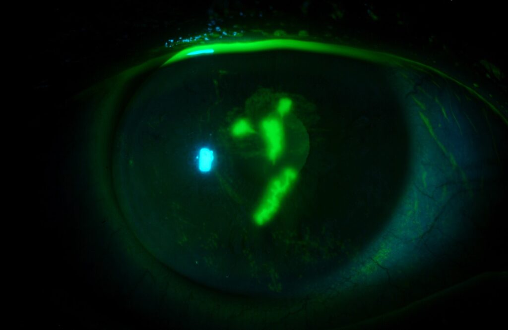
Acanthamoeba Sampling
Acanthamoeba cysts may appear on calcofluor white stains.
Also, confocal microscopy can show cysts at the bedside.
Transport and Handling
Plate the sample at the bedside if possible.
Also, send slides and swabs to the laboratory without delay.
Because delays reduce yield, label samples with the suspected organism.
Record Keeping After Corneal Scraping
Note the ulcer size, depth, infiltrate and hypopyon in the chart.
Also, photograph the cornea at the slit lamp.
Because response is judged by change, photographs help follow the course.
Antibiotic Sensitivity
Culture results with sensitivities guide the choice of drops.
Also, interpret breakpoints with caution, since they derive from systemic drug levels while topical drops reach very high concentrations.
Therefore, clinical response matters as much as laboratory results.
Corneal Scraping in Resource-Limited Settings
A Gram stain and a potassium hydroxide preparation give rapid results.
Also, training staff to take good samples improves care.
Instrument Choices
A Bard-Parker blade, a Kimura spatula and a sterile needle are common choices.
Also, a calcium alginate swab can collect material from a small ulcer.
Because each tool has a trade-off, pick the one you can control best.
Training and Simulation
Practice on a model or a fruit before you work on a patient.
Also, observe an experienced colleague and then perform scrapings under supervision.
Therefore, confidence and sample quality improve over time.
Pitfalls That Reduce Yield
Antibiotic drops given before the sample reduce growth.
Using a small amount of anesthetic with preservatives also lowers yield.
Also, scraping only the surface of a deep ulcer misses the organism.
Because a good sample guides therapy, take it before treatment whenever possible.
Then record which media and slides you prepared.


Document what you see
Two smartphone imaging tools built for everyday clinic use — one for the slit lamp, one for the fundus.
From Choroida — the team behind this siteReferences
- Lin A, Rhee MK, Akpek EK, et al. Bacterial Keratitis Preferred Practice Pattern. Ophthalmology. 2019;126:P1-P55.
- McLeod SD, Kolahdouz-Isfahani A, Rostamian K, Flowers CW, Lee PP, McDonnell PJ. The role of smears, cultures, and antibiotic sensitivity testing in the management of suspected infectious keratitis. Ophthalmology. 1996;103:23-28.
- Srinivasan M, Gonzales CA, George C, et al. Epidemiology and aetiological diagnosis of corneal ulceration in Madurai, south India. Br J Ophthalmol. 1997;81:965-971.
- Leck A, Burton M. Distinguishing fungal and bacterial keratitis on clinical signs. Community Eye Health. 2015;28:6-7.

Writes and reviews the clinical articles on Educate. Every post is checked against peer-reviewed sources before it goes live.