The foveal avascular zone, or FAZ, is the central macular area that has no retinal capillaries.
It keeps the fovea clear for light to reach the photoreceptors, and its size and shape vary widely between healthy people.
OCT angiography now allows detailed measurement, and changes in the FAZ signal disease.
What the Foveal Avascular Zone Is
The FAZ lies at the center of the fovea, and the capillaries around it form a ring.
It is roughly circular, with an average diameter around 0.5 mm, although normal values vary.
It is larger in people with a thin fovea, and it differs between the superficial and deep capillary plexuses.
Bresnick and colleagues described its enlargement in diabetic retinopathy using fluorescein angiography.
How to Measure the Foveal Avascular Zone
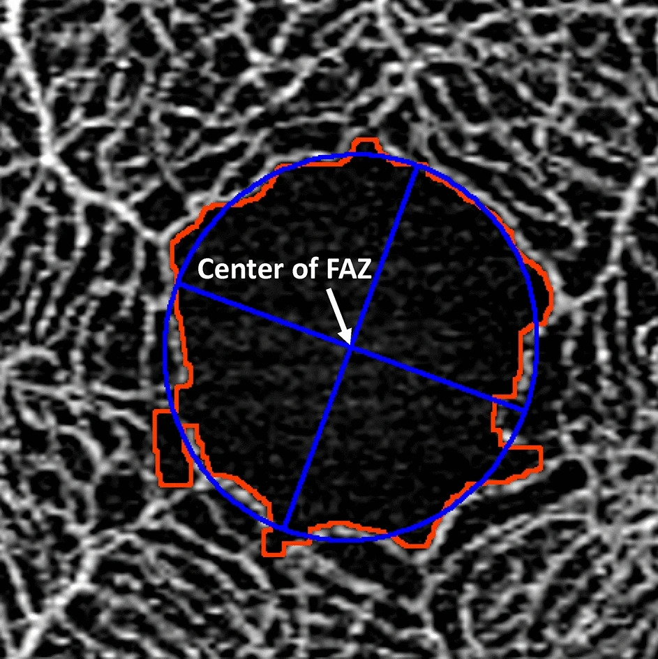
OCT angiography gives a clear en face image of the capillary plexuses.
Software outlines the FAZ on the superficial slab, and it calculates the area, the perimeter, and the circularity.
Manual correction improves accuracy when the software fails (see OCT angiography).
Linderman and colleagues showed that the metrics vary between devices and with scan settings.
Fundus Explorer Pro
Photograph the retinal findings described here with the phone already in your pocket — 22 D optics and built-in illumination in one handheld unit.
From Choroida — the team behind this siteFoveal Avascular Zone in Diabetic Retinopathy
In diabetes, the FAZ enlarges and becomes irregular as capillaries drop out near the fovea.
Samara and colleagues found that FAZ enlargement correlates with vision and with the severity of retinopathy.
Therefore, it marks macular ischemia, which may limit vision gains (see diabetic macular ischemia).
Other Conditions That Change the Foveal Avascular Zone
- Retinal vein occlusion, with enlargement from capillary loss
- Sickle cell retinopathy, with temporal enlargement and thinning
- Retinopathy of prematurity and foveal hypoplasia, with a small or absent FAZ
- Glaucoma and Alzheimer disease, with changes reported in some studies
A small or absent FAZ suggests abnormal foveal development (see albinism and foveal hypoplasia).
Pitfalls in Measuring the Foveal Avascular Zone
- Using different devices or settings between visits
- Ignoring signal strength and motion artifacts
- Reading FAZ area without checking the B-scan and segmentation
- Comparing patients without adjusting for refraction and axial length
Axial length changes scaling, so adjust measurements in myopic eyes.
Clinical Use of the Foveal Avascular Zone
Use FAZ metrics to monitor disease and to support counseling.
Record the area at baseline and compare with follow-up.
Treat the numbers as supporting data, since normal ranges are wide.
An Example Case
A 52-year-old man with type 2 diabetes has mild nonproliferative retinopathy and good vision.
OCT angiography shows an enlarged and irregular foveal avascular zone in the right eye, with areas of capillary dropout.
The finding raises concern for macular ischemia, so the clinician shortens the review interval and reinforces glycemic control.
Over two years, the zone enlarges further, and the retinopathy progresses.
Questions Patients Ask
Patients ask whether the zone is a problem, and the answer is that some variation is normal.
They also ask whether the size predicts vision, and a larger zone often goes with worse vision in disease.
Because values vary, explain that follow-up measures matter more than a single number.
Pitfalls to Avoid
- Judging the zone by the automatic outline without checking it
- Mixing measurements from different devices
- Comparing eyes with different axial lengths without adjustment
- Overinterpreting small differences in area
Looking Ahead
Standardized software and normative databases will make measurements more reliable.
Also, deep learning may segment the zone with less error.
Therefore, the zone may become a routine marker of retinal ischemia.
Practical Points
Use the same device, scan size, and slab each time.
Then confirm the segmentation before recording the area.
Also note the signal strength with each measurement.
Normal Variation
The zone differs by age, sex, axial length, and central macular thickness.
Also, it appears smaller in the deep plexus than in the superficial plexus in many eyes.
Because of this variation, a single measurement rarely makes a diagnosis.
Instead, use the shape and the surrounding capillaries to judge disease.
Reporting Metrics
Report area, perimeter, and circularity, and note the device and slab used.
Then compare changes over time with the same settings.
Also describe the vessels around the zone, such as dropout or loops.
Check that the scan centers on the fovea, since off-center scans distort the zone.
Because patient movement shifts the image, repeat poor scans.
Clinical Scenarios
A patient with diabetes and a large zone needs closer follow-up.
Also, a patient with vein occlusion and an enlarged zone may have a poorer visual outlook.
Because the zone reflects ischemia, discuss risk factors such as blood pressure and glucose.
Then plan the next scan at a fixed interval.
Therefore, the measurement helps to personalize care.
For the latest studies, see the PubMed results for foveal avascular zone.


Document what you see
Two smartphone imaging tools built for everyday clinic use — one for the slit lamp, one for the fundus.
From Choroida — the team behind this siteReferences
- Bresnick GH, Condit R, Syrjala S, et al. Abnormalities of the foveal avascular zone in diabetic retinopathy. Arch Ophthalmol. 1984;102:1286-1293.
- Samara WA, Shahlaee A, Adam MK, et al. Quantification of diabetic macular ischemia using optical coherence tomography angiography and its relationship with visual acuity. Ophthalmology. 2017;124:235-244.
- Linderman RE, Muthiah MN, Omoba SB, et al. Variability of foveal avascular zone metrics derived from optical coherence tomography angiography images. Transl Vis Sci Technol. 2018;7:20.
- American Academy of Ophthalmology. Basic and Clinical Science Course, Section 12: Retina and Vitreous. AAO; current edition.

Writes and reviews the clinical articles on Educate. Every post is checked against peer-reviewed sources before it goes live.