
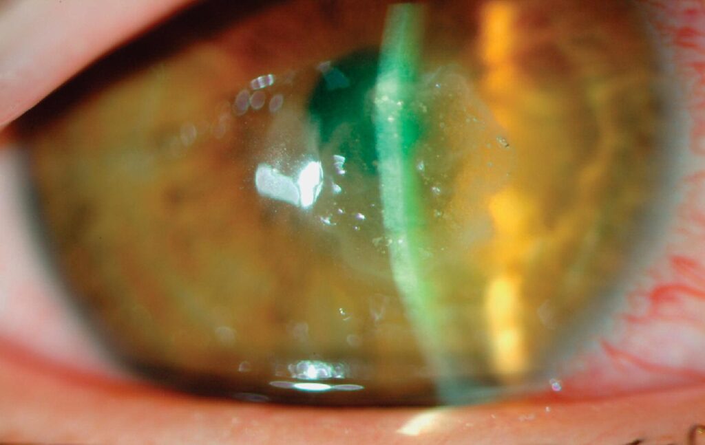
Nocardia keratitis is an uncommon corneal infection caused by aerobic actinomycetes of the genus Nocardia, and it often follows trauma with vegetable matter or soil.
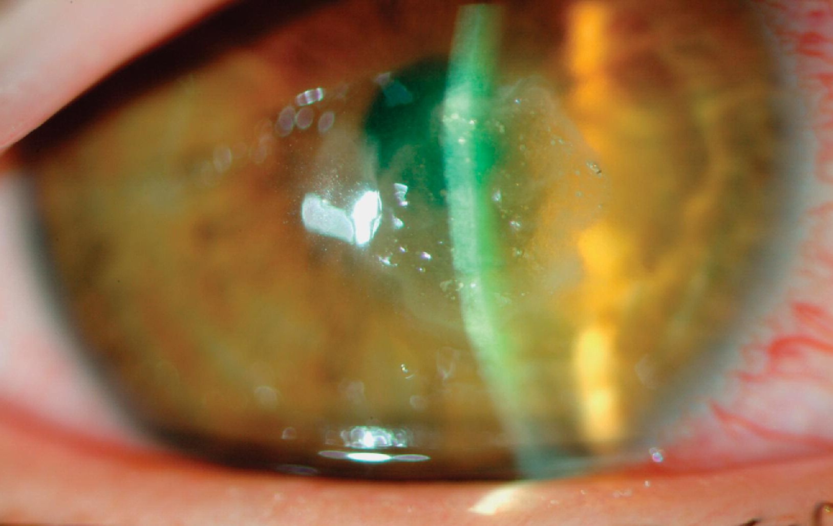
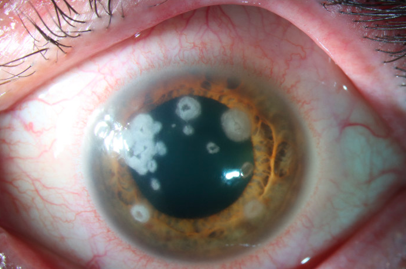
The ulcer progresses slowly, and its appearance resembles a fungal infection, which leads to frequent misdiagnosis and delayed treatment.
The diagnosis depends on laboratory confirmation, and the treatment differs from that of the more common bacterial and fungal ulcers.
What Is Nocardia Keratitis?
Nocardia are gram-positive, weakly acid-fast, branching filamentous bacteria that live in soil and water.
They enter the cornea through an abrasion or a penetrating injury, or after surgery or contact lens use.
Most cases occur in agricultural settings or after injury with plants, and a proportion follows refractive or other corneal surgery.
Nocardia asteroides complex and Nocardia brasiliensis are among the species reported in corneal infection.
Clinical Features
The patient has pain, redness, tearing, and reduced vision, and the onset is subacute and not abrupt.
The infiltrate is gray-white, often with a dry, granular surface and a wreath-like pattern of small, discrete, chalky patches in the anterior stroma.
Fine radiating lines or “cracked windshield” patterns can be present.
Satellite lesions and feathery margins suggest a fungus, and this is the main source of confusion.
Hypopyon can occur in advanced disease, and the infiltrate may extend deeper over time.
All-fit Slit-Lamp Adapter
Record and share exactly what you see at the slit lamp. One adapter fits any slit lamp or surgical microscope — and any smartphone.
From Choroida — the team behind this siteDiagnosis
Corneal scraping for Gram stain, acid-fast stain, and culture is the basis of diagnosis.
Gram stain shows beaded, branching gram-positive rods, and a modified acid-fast stain, using a weak acid for decolorization, can help to separate Nocardia from other actinomycetes.
Culture on blood agar and other media, with prolonged incubation, produces dry, chalky colonies.
The laboratory should be told that Nocardia is suspected, because it can be overlooked or discarded as a contaminant.
Susceptibility testing is useful, since resistance patterns vary among species.
Differential Diagnosis
- Fungal keratitis: feathery edges and satellites, typically with a history of plant trauma (see fungal versus bacterial keratitis)
- Nontuberculous mycobacteria: a slow, indolent ulcer with a ground-glass appearance
- Herpes simplex keratitis: reduced sensation and a response to antivirals
- Acanthamoeba keratitis: pain out of proportion and a ring infiltrate (see Acanthamoeba keratitis)
A lack of response to antifungal and standard antibacterial therapy, with a negative fungal culture, should raise the question of Nocardia (see why keratitis fails treatment).
Treatment
Topical amikacin is the standard first-line agent, and it is used as a fortified drop at frequent intervals.
Trimethoprim-sulfamethoxazole and other sulfonamides are alternatives, and some patients need both.
Oral treatment is added for deep infection, and therapy usually continues for weeks.
Debridement of the necrotic tissue may speed healing, and therapeutic keratoplasty is reserved for perforation or ulcers that fail medical therapy.
Steroids are avoided until the infection is controlled, and the patient needs frequent review because of the slow response.
Why It Is Often Mistaken for Fungal Keratitis
Nocardia grows as branching filaments, so a smear can show structures that resemble fungal hyphae, and the infiltrate has the feathery edge and dry, raised surface that are seen in fungal ulcers.
The clinical history is similar, since many patients have an injury in a rural setting, and the course is slow.
The mistake is easy to make, and it is only corrected when the culture grows an organism that does not match a fungus, or when the ulcer fails to respond to antifungal agents.
Because both conditions are slow, a small delay is not disastrous, but a long delay allows deep stromal involvement.
Microbiology Tips
Ask the laboratory to hold the plates for at least two weeks, because growth may be slow.
A modified Kinyoun or Ziehl-Neelsen stain with a weak acid decolorizer helps, and the gram stain of a scraping in an ulcer with a wreath-like appearance should be examined carefully for beaded filaments.
If only a small sample can be obtained, prioritize culture and smear over other tests.
Patient Counselling
Explain that this infection is slow and that recovery is also slow, often over weeks to months.
The drops are used frequently at first and then tapered, and the patient should expect a scar at the site.
Advise protective eyewear for work in fields or gardens, and prompt treatment of any corneal injury from plants or soil.
Keep in mind that Nocardia infection can also affect immunocompromised patients systemically, so a patient with a positive culture and a history of immunosuppression, lung disease, or skin lesions deserves a medical review.
The corneal infection itself is local in most cases.


Document what you see
Two smartphone imaging tools built for everyday clinic use — one for the slit lamp, one for the fundus.
From Choroida — the team behind this siteReferences
- Lalitha P, Tiwari M, Prajna NV, et al. Nocardia keratitis: species, drug sensitivities, and clinical correlation. Cornea. 2007;26:255-259.
- Lalitha P, Srinivasan M, Rajaraman R, et al. Nocardia keratitis: clinical course and effect of corticosteroids. Am J Ophthalmol. 2012;154:934-939.
- Mannis MJ, Holland EJ, eds. Cornea. 4th ed. Elsevier; 2017.
- American Academy of Ophthalmology. Basic and Clinical Science Course, Section 8: External Disease and Cornea. AAO; current edition.

Writes and reviews the clinical articles on Educate. Every post is checked against peer-reviewed sources before it goes live.


