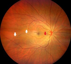
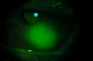
Corneal infiltrates are white or gray stromal opacities formed by inflammatory cells that migrate into the cornea.
Some reflect active infection, while others are an immune response to bacterial antigens, lens wear or a virus.
The bedside task is to decide which is which at the first visit.
Delay in infectious keratitis costs vision, whereas overtreating a sterile lesion adds toxicity.
What Corneal Infiltrates Are
Leukocytes reach the cornea from limbal vessels and the tear film.
They accumulate in the anterior stroma and scatter light, so the area looks white.
An infiltrate does not prove infection by itself.
Therefore, the surrounding signs carry most of the diagnostic weight.
History That Shifts the Odds
Start with a few targeted questions.
- Contact lens wear, especially overnight or with tap water exposure
- Trauma with vegetable matter
- Recent conjunctivitis or a sick contact
- Blepharitis, rosacea or previous similar episodes
- Topical steroid use or recent ocular surgery
All-fit Slit-Lamp Adapter
Record and share exactly what you see at the slit lamp. One adapter fits any slit lamp or surgical microscope — and any smartphone.
From Choroida — the team behind this sitePain and Corneal Infiltrates
Infectious lesions usually hurt more than their size suggests.
Sterile marginal infiltrates often cause mild irritation and redness only.
However, severe pain with minimal findings should raise suspicion of Acanthamoeba.
Also remember that reduced sensation in herpetic or neurotrophic eyes can mask pain.
Location at the Slit Lamp
Central and paracentral lesions are more likely infectious.
In contrast, sterile infiltrates tend to sit in the periphery.
Staphylococcal marginal keratitis classically leaves a clear zone between infiltrate and limbus.
This lucid interval appears because immune complexes deposit near limbal vessels.
Epithelium Over Corneal Infiltrates
Fluorescein staining is the most useful single test.
An infectious ulcer usually shows an epithelial defect matching or exceeding the infiltrate.
Sterile infiltrates often have intact epithelium or a defect smaller than the opacity.
However, a sterile lesion can break down, so recheck staining within one to two days.
Size, Number and Shape
Small, multiple, round lesions favor a sterile process.
A single large infiltrate with indistinct, suppurative edges favors infection.
Feathery borders and satellite lesions also suggest fungal keratitis.
Then, a ring infiltrate with radial perineuritis points toward Acanthamoeba.
Anterior Chamber Reaction
Look carefully for cells, flare and hypopyon.
A sterile infiltrate rarely produces more than a trace reaction.
Therefore, treat a moderate reaction or any hypopyon as infection until proven otherwise.
Contact Lens Corneal Infiltrates
Soft lens wearers develop a spectrum of infiltrative events.
Peripheral ulcers in lens wearers are small, round and often leave a scar.
Acute red eye from lens wear presents on waking with diffuse small infiltrates.
However, microbial keratitis can start with the same small lesion, so never dismiss lens wearers casually.
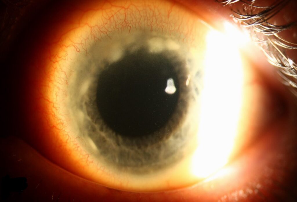
Viral Subepithelial Infiltrates
Adenoviral keratoconjunctivitis can leave multiple subepithelial infiltrates one to two weeks after onset.
They lie under intact epithelium across the central cornea.
Patients report glare and blur more than pain.
In addition, a preceding red, watery eye usually clarifies the diagnosis.
The 1, 2, 3 Rule for Corneal Infiltrates
Vital and colleagues proposed a simple triage rule.
Culture when the anterior chamber shows at least 1+ cells, the infiltrate measures 2 mm or more, or it lies within 3 mm of the center.
Lesions outside these limits can usually receive empirical therapy.
Still, atypical features justify scraping.
When to Scrape and Culture
The AAO Preferred Practice Pattern recommends cultures in specific settings.
- Central, large or deep infiltrates
- Chronic lesions or failure to respond to broad-spectrum therapy
- Atypical features suggesting fungus, Acanthamoeba or mycobacteria
- Prior corneal surgery
- Multifocal infiltrates
Scrape the base and edge, then inoculate media and prepare smears.
Empirical Treatment of Corneal Infiltrates
Small peripheral lesions without red flags may start on a topical fluoroquinolone.
Fortified antibiotics remain the usual choice for large or central ulcers.
Also record slit-beam dimensions to track the response objectively.
Review the patient within 24 to 48 hours.
Steroids in Sterile Infiltrates
Marginal keratitis responds quickly to a short course of topical steroid.
However, steroids worsen fungal and herpetic disease.
In culture-proven bacterial keratitis, the SCUT trial found no overall difference in visual acuity at three months with adjunctive steroids.
Therefore, start them only once infection is reasonably excluded or controlled.
Follow-Up of Corneal Infiltrates
A sterile lesion should improve within days.
Enlargement, new epithelial breakdown or thinning suggests infection or another process.
Peripheral melting with scleritis may reflect systemic disease (see sterile corneal melt).
Recurrent marginal disease may also leave vessels behind (see corneal pannus).
Counseling the Patient
Explain that a white spot can be inflammation or infection, and early review decides the plan.
Ask lens wearers to stop wearing lenses and bring the case.
Then give clear return precautions for worsening pain, discharge or vision.
Pitfalls to Avoid
- Do not start steroids before excluding an epithelial defect
- Never assume a lens wearer’s small infiltrate is sterile without review
- Do not overlook herpes when corneal sensation is reduced
For the latest studies, see the PubMed results for corneal infiltrates.


Document what you see
Two smartphone imaging tools built for everyday clinic use — one for the slit lamp, one for the fundus.
From Choroida — the team behind this siteReferences
- Vital MC, Belloso M, Prager TC, Lanier JD. Classifying the severity of corneal ulcers by using the “1, 2, 3” rule. Cornea. 2007;26:16-20.
- Lin A, Rhee MK, Akpek EK, et al. Bacterial Keratitis Preferred Practice Pattern. Ophthalmology. 2019;126:P1-P55.
- Srinivasan M, Mascarenhas J, Rajaraman R, et al. Corticosteroids for bacterial keratitis: the Steroids for Corneal Ulcers Trial (SCUT). Arch Ophthalmol. 2012;130:143-150.
- Sweeney DF, Jalbert I, Covey M, et al. Clinical characterization of corneal infiltrative events observed with soft contact lens wear. Cornea. 2003;22:435-442.

Writes and reviews the clinical articles on Educate. Every post is checked against peer-reviewed sources before it goes live.