Pseudodendrite is a raised, branching epithelial lesion of the cornea, most often seen in herpes zoster ophthalmicus.
At first glance, it can mimic the dendritic ulcer of herpes simplex virus. However, its morphology, staining and treatment differ in ways you can see at the slit lamp.
What a Pseudodendrite Is
A pseudodendrite forms from swollen, heaped-up epithelial cells, and there is no true ulcer because the epithelium stays largely intact. As a result, the lesion sits slightly above the corneal surface, and varicella zoster virus is the classic cause.
When Zoster Pseudodendrites Appear
Zoster epithelial disease often starts as punctate epithelial keratitis within the first days of the rash, and these spots may then coalesce into pseudodendrites. Later, delayed mucous plaques can appear with a similar look, so timing relative to the rash helps classify the lesion (see herpes zoster ophthalmicus).
All-fit Slit-Lamp Adapter
Record and share exactly what you see at the slit lamp. One adapter fits any slit lamp or surgical microscope — and any smartphone.
From Choroida — the team behind this sitePseudodendrite Morphology at the Slit Lamp
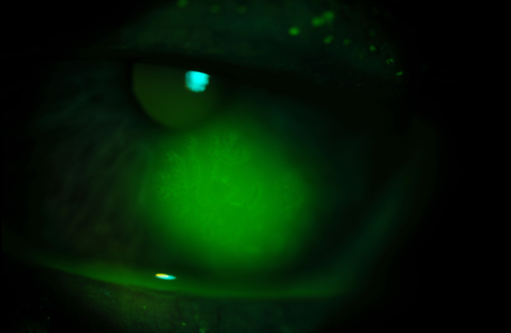
The lesion looks coarse, ropy and gray. Its branches are short, thick and irregular, sometimes described as medusa-like, and the arms taper and end without any swelling.
The True HSV Dendrite
An HSV dendrite is a genuine epithelial ulcer with a fine, branching pattern, and its delicate branches divide repeatedly. Each branch typically ends in a terminal bulb, and swollen, virus-laden cells line the ulcer margin.
Terminal Bulbs and Branch Pattern
Terminal end bulbs are the most reliable bedside discriminator because they reflect active viral spread at the ulcer tips. In contrast, a pseudodendrite lacks them, so inspect every branch tip under high magnification.
Fluorescein Staining in Pseudodendrite
Fluorescein fills the base of a true dendritic ulcer and stains it brightly. A pseudodendrite, however, stains faintly or patchily, and the dye may instead pool around its raised edges.
Rose Bengal and Lissamine Green
These dyes stain devitalized and swollen epithelial cells, so they highlight, for example, the margins of an HSV dendrite. They also stain zoster pseudodendrites well because the lesion consists of abnormal cells, so combine dyes when the picture is unclear (see corneal staining patterns).
Corneal Sensation and Associated Signs
Both viruses can reduce corneal sensation, but zoster often causes a deeper and more lasting loss. Check the forehead, lids and nose tip for vesicles or scars, and also look for stromal infiltrates, keratic precipitates and sectoral iris atrophy (see viral anterior uveitis).
History That Shifts the Diagnosis
A recent painful dermatomal rash points strongly to zoster, while recurrent unilateral episodes and cold sores favor HSV, although VZV occasionally causes epithelial disease without a rash.
Laboratory Confirmation of a Pseudodendrite
Most cases need only a clinical diagnosis. However, PCR of an epithelial swab can separate VZV from HSV when findings are atypical, while viral culture is slower and less sensitive.
Other Causes of Pseudodendrites
- Early epithelial Acanthamoeba keratitis
- Healing corneal abrasion with irregular epithelial ridges
- Contact lens related epitheliopathy
- Topical medication toxicity
- Tyrosinemia type II
Treating a Zoster Pseudodendrite
Oral antiviral therapy for zoster is the main treatment, and acyclovir, valacyclovir or famciclovir started early can reduce ocular complications. Pseudodendrites usually fade as the acute phase settles. Some clinicians add topical ganciclovir gel, although evidence for this remains limited, and debridement has no established role here.
Treating an HSV Dendrite
HSV epithelial keratitis responds to topical or oral antivirals, with options including ganciclovir gel, trifluridine drops, oral acyclovir or valacyclovir. Gentle debridement can also speed healing alongside an antiviral.
Why Steroids Need Caution
Topical steroids can enlarge an HSV dendrite into a geographic ulcer, so avoid them in active HSV epithelial disease. Zoster patients may still need steroids for stromal keratitis or uveitis, but confirm the diagnosis first, because a misread dendrite carries real risk.
Follow-Up in Pseudodendrite Cases
Review the patient within a few days, then watch for stromal keratitis, uveitis, raised pressure and neurotrophic changes. Zoster eye disease can also recur months or years later.
Counseling the Patient
Explain that the lesion is a surface sign of viral eye disease, and first stress completing the full oral antiviral course. Next, warn against using leftover steroid drops, and finally discuss shingles vaccination after recovery.
Pitfalls to Avoid
- Do not label every branching lesion as HSV before checking the branch tips
- Never start topical steroids until you exclude an HSV dendrite
- Do not skip lissamine green when fluorescein staining looks faint
- Consider Acanthamoeba in contact lens wearers


Document what you see
Two smartphone imaging tools built for everyday clinic use — one for the slit lamp, one for the fundus.
From Choroida — the team behind this siteReferences
- Liesegang TJ. Herpes zoster ophthalmicus natural history, risk factors, clinical presentation, and morbidity. Ophthalmology. 2008;115(2 Suppl):S3-S12.
- Cobo LM, Foulks GN, Liesegang T, et al. Oral acyclovir in the treatment of acute herpes zoster ophthalmicus. Ophthalmology. 1986;93:763-770.
- Cohen EJ, Kessler J. Persistent dilemmas in zoster eye disease. Br J Ophthalmol. 2016;100:56-61.
- Wilhelmus KR. Antiviral treatment and other therapeutic interventions for herpes simplex virus epithelial keratitis. Cochrane Database Syst Rev. 2015;1:CD002898.
- American Academy of Ophthalmology. Basic and Clinical Science Course, Section 8: External Disease and Cornea. San Francisco: AAO; 2023.
Test yourself
A few questions straight from this article.
-
What forms a zoster corneal pseudodendrite?
A pseudodendrite forms from swollen, heaped-up epithelial cells; there is no true ulcer, so the lesion sits slightly above the corneal surface. -
How does zoster epithelial disease often begin within the first days of the rash?
Punctate epithelial keratitis often appears first and may coalesce into pseudodendrites, while mucous plaques can appear later with a similar look. -
Which branch pattern describes a zoster pseudodendrite at the slit lamp?
The pseudodendrite looks coarse, ropy and gray with short, thick, irregular branches, sometimes described as medusa-like, and its arms taper without swelling. -
What is the most reliable bedside discriminator of an HSV dendrite from a zoster pseudodendrite?
Terminal end bulbs reflect active viral spread at the ulcer tips and are absent from a pseudodendrite, so every branch tip should be inspected under high magnification. -
How does a zoster pseudodendrite usually behave with fluorescein compared with a true HSV dendrite?
Fluorescein fills the base of a true dendritic ulcer and stains it brightly, whereas a pseudodendrite stains faintly or patchily and dye may pool around its raised edges. -
A suspected zoster pseudodendrite stains faintly with fluorescein. Which dye should be added?
Rose bengal and lissamine green stain devitalized and swollen epithelial cells, and they stain zoster pseudodendrites well because the lesion consists of abnormal cells. -
What is the main treatment for a zoster pseudodendrite?
Oral antiviral therapy for zoster is the main treatment, and acyclovir, valacyclovir or famciclovir started early can reduce ocular complications. -
What can topical steroids do to an active HSV dendrite?
Topical steroids can enlarge an HSV dendrite into a geographic ulcer, so they are avoided in active HSV epithelial disease and the diagnosis is confirmed first. -
What role does debridement have in managing a zoster pseudodendrite?
Debridement has no established role for zoster pseudodendrites, whereas gentle debridement can speed healing of an HSV dendrite alongside an antiviral. -
Which test can separate VZV from HSV when a branching corneal lesion looks atypical?
Most cases need only a clinical diagnosis, but PCR of an epithelial swab can separate VZV from HSV when findings are atypical; viral culture is slower and less sensitive.

Writes and reviews the clinical articles on Educate. Every post is checked against peer-reviewed sources before it goes live.