
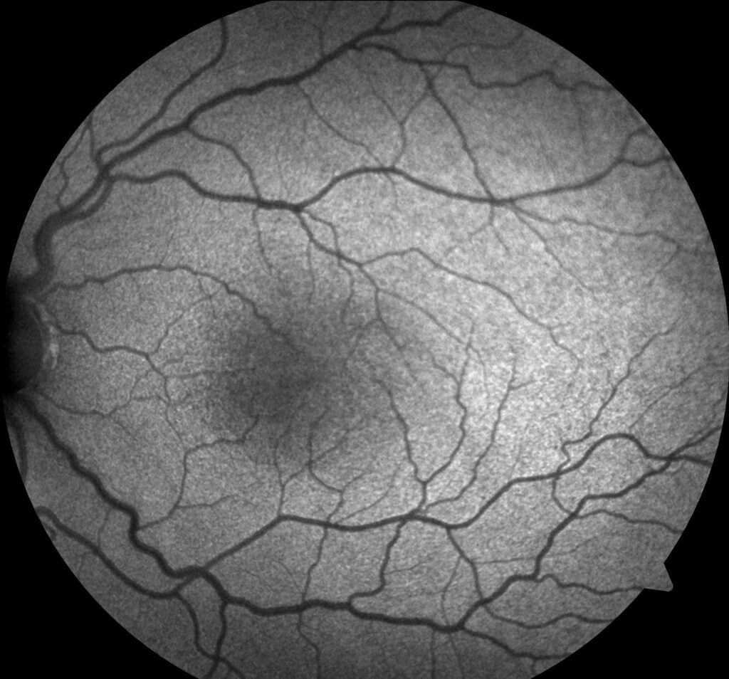
Foveal hypoplasia in albinism is the structural finding that best explains why children with albinism have reduced vision, and it matters more for predicting visual outcome than the degree of iris or fundus pigmentation, which varies widely and correlates poorly with acuity.
OCT has made it possible to grade this finding precisely, turning what used to be a qualitative fundus impression into a reproducible measurement.
Why the Fovea Fails to Develop Normally
The normal fovea develops through a process of centrifugal displacement of the inner retinal layers, concentration of cone photoreceptors, and formation of a foveal pit, continuing after birth into early childhood.
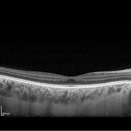
In albinism, reduced or absent melanin during this developmental window disrupts the signaling that drives normal foveal maturation, leaving a fovea that lacks its normal pit, has a thinner outer nuclear layer than expected, and often shows persistence of inner retinal layers that should have migrated away from the center.
OCT Grading of Foveal Hypoplasia
A widely used grading system classifies foveal hypoplasia by the presence or absence of specific structural features on OCT, arranged in increasing severity.
- Grade 1: a shallow foveal pit is present, with normal outer retinal layers but incomplete lamination
- Grade 2: absence of the foveal pit, with continued presence of inner retinal layers across the fovea
- Grade 3: absence of the foveal pit with additional outer segment lengthening abnormalities
- Grade 4: absence of both the foveal pit and the normal extrusion of the outer plexiform layer, representing the most severe grade
More severe grades correlate with worse visual acuity, giving OCT grading real prognostic value beyond what fundus examination alone provides.
Fundus Explorer Pro
Photograph the retinal findings described here with the phone already in your pocket — 22 D optics and built-in illumination in one handheld unit.
From Choroida — the team behind this siteAssociated Ocular Findings in Albinism
- Nystagmus, usually present from infancy and often the presenting sign that brings the child to attention
- Iris transillumination, from reduced iris pigment epithelium
- Fundus hypopigmentation, with visible large choroidal vessels
- Chiasmal misrouting of the optic nerve fibers, detectable on visual evoked potential testing, a feature specific to albinism among causes of infantile nystagmus
- Strabismus and reduced stereopsis, related to the abnormal chiasmal decussation
Types of Albinism
Albinism includes oculocutaneous forms, affecting skin, hair, and eyes, and ocular albinism, largely confined to the eyes with normal or near-normal skin and hair pigmentation (see ocular albinism).
Multiple genes are implicated across the different types, and genetic testing can confirm the specific subtype, which is useful for genetic counseling even though it does not usually change ophthalmic management.
Diagnostic Value of OCT
Before OCT was widely available, the diagnosis of albinism-related visual impairment relied on fundus appearance, iris transillumination, and VEP chiasmal misrouting testing.
OCT now provides a fast, noninvasive, and highly informative structural assessment, and it can be performed even in young children with handheld OCT devices, making it useful both for confirming the diagnosis and for counseling families about expected visual potential.
Foveal hypoplasia is not unique to albinism and is also seen in aniridia, achromatopsia, and some other conditions, so OCT findings are interpreted alongside the rest of the clinical picture (see aniridia).
Management
There is no treatment that restores normal foveal architecture, so management centers on optimizing the vision that is present.
- Refractive correction, since significant refractive errors are common
- Management of nystagmus and strabismus as needed
- Low-vision support and magnification, tailored to the degree of visual impairment
- Sun protection and skin cancer surveillance in oculocutaneous forms, coordinated with dermatology
Prognosis
Visual acuity in albinism varies widely, generally correlating with the severity of foveal hypoplasia on OCT more closely than with the degree of pigment loss.
Most children achieve functional vision adequate for mainstream education with appropriate correction and support, even though acuity typically remains below normal throughout life.


Document what you see
Two smartphone imaging tools built for everyday clinic use — one for the slit lamp, one for the fundus.
From Choroida — the team behind this siteReferences
- Thomas MG, Kumar A, Mohammad S, et al. Structural grading of foveal hypoplasia using spectral-domain optical coherence tomography: a predictor of visual acuity? Ophthalmology. 2011;118:1653-1660.
- Mohammad S, Gottlob I, Kumar A, et al. The functional significance of foveal abnormalities in albinism measured using spectral-domain optical coherence tomography. Ophthalmology. 2011;118:1645-1652.
- Grønskov K, Ek J, Brondum-Nielsen K. Oculocutaneous albinism. Orphanet J Rare Dis. 2007;2:43.
- McCafferty BK, Wilk MA, McAllister JT, et al. Clinical insights into foveal morphology in albinism. J Pediatr Ophthalmol Strabismus. 2015;52:167-172.
Test yourself
A few questions straight from this article.
-
In albinism, which finding correlates most closely with visual acuity?
Iris and fundus pigmentation vary widely and correlate poorly with acuity, whereas OCT-graded foveal hypoplasia carries real prognostic value. -
What disrupts normal foveal maturation in albinism?
Reduced or absent melanin disrupts the signalling that drives foveal maturation, leaving no pit, a thinner outer nuclear layer and persisting inner retinal layers. -
On OCT grading of foveal hypoplasia, what characterises grade 2?
Grade 2 is defined by absence of the foveal pit together with continued presence of inner retinal layers across the fovea. -
What makes OCT feasible for assessing foveal hypoplasia in very young children?
Handheld OCT allows a fast, noninvasive structural assessment even in young children, helping both confirm the diagnosis and counsel families. -
Which feature is specific to albinism among causes of infantile nystagmus?
Chiasmal misrouting of the optic nerve fibres, detectable on VEP testing, is specific to albinism among causes of infantile nystagmus. -
Iris transillumination in albinism reflects reduced pigment in which structure?
Reduced iris pigment epithelium allows light to pass through the iris, producing the transillumination seen on slit lamp examination. -
Besides albinism, foveal hypoplasia is a recognised feature of which conditions?
Foveal hypoplasia is not unique to albinism; aniridia and achromatopsia are among the other causes, so OCT is read alongside the clinical picture. -
What distinguishes ocular albinism from the oculocutaneous forms?
Ocular albinism is largely confined to the eyes, with normal or near-normal skin and hair pigmentation, unlike the oculocutaneous forms. -
Which extra-ocular surveillance is part of managing oculocutaneous albinism?
Sun protection and skin cancer surveillance, coordinated with dermatology, form part of management in the oculocutaneous forms. -
What is the usual visual and educational outlook for children with albinism?
With appropriate correction and support most children reach vision sufficient for mainstream education, although acuity typically stays below normal for life.

Writes and reviews the clinical articles on Educate. Every post is checked against peer-reviewed sources before it goes live.


