
Introduction
It is evident from the name that it is a macular disease, So first you should know what a macula is. The central part of the retina which provides central, sharp, clear, and color vision is called the macula.
This area contains special photoreceptor cells which are sensitive to light. These photoreceptor cells receive light and color signals to make you capable of seeing objects colorful and bright.
DISEASE
Diabetic Macular ischemia is a decrease in blood flow to the macula due to uncontrolled diabetes.
In diabetes, continuous uncontrolled blood glucose level affects the retinal microvessels thus causing either leakage or occlusion of capillaries and pre-capillary arterioles.

Leakage of retinal capillaries:
Retinal capillaries are lined by a single membrane of endothelium and overlying pericytes which are the supporting cells of capillaries and allow the selective matter to diffuse out of the blood.
Due to hyperglycemia, pericytes are lost and the junctions between endothelial cells weaken, so the capillary wall becomes abnormally permeable and allows the vascular content to leak out thus causing microaneurysms, hard exudates and hemorrhages, and retinal edema.
Occlusion of Retinal capillaries:
Raised blood glucose level damages the capillary wall and causes their basement membrane to thicken. Due to their thickening, firstly microvessels become narrow and then get blocked completely.
This Occlusion does not allow the blood to reach the retina including the macula thus devoiding it of oxygen and nutrition and making the Macular region ischemic.
It is an associated Complication of diabetic retinopathy (DR) and vision loss caused by it is irreversible.
Fundus Explorer Pro
Photograph the retinal findings described here with the phone already in your pocket — 22 D optics and built-in illumination in one handheld unit.
From Choroida — the team behind this siteDiabetic Macular ischemia Diagnostic tests
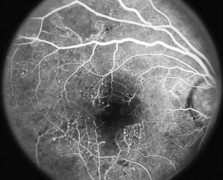
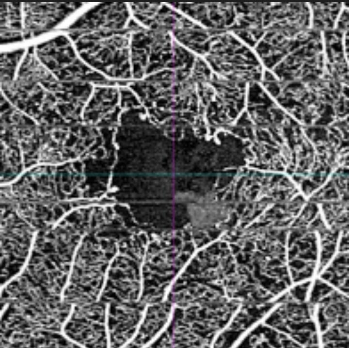
The following test can be done to diagnose DMI
- OCTA (Optical Coherence Tomography Angiography)
This new technique is useful in visualizing the layers of retinal capillaries without injecting fluorescein sodium dye
- FA (Fluorescein Angiography)
Symptoms
Decreased visual acuity, which is often gradual and may go unnoticed early on.
Diabetic Macular ischemia MANAGEMENT
Until now, there is no defined treatment for DMI. But managing the risk factors like controlling blood glucose level and blood pressure can prevent developing DMI. Anti-VEGF and steroids have brought no significant improvement in treating DMI
Laser photocoagulation is used for the associated diabetic retinopathy and macular edema, but it does not reverse macular ischemia and carries a risk of complications.
As this disease may cause no noticeable symptoms early on, you should regularly monitor your glucose level and blood pressure. As soon as you notice a regular rise in BP or hyperglycemia you should consult an eye doctor for a detailed fundus examination and continue follow-up visits from time to time to be on the safer side.
Risk factors
High blood glucose levels and high blood pressure are contributing risk factors for Diabetic Macular ischemia.


Document what you see
Two smartphone imaging tools built for everyday clinic use — one for the slit lamp, one for the fundus.
From Choroida — the team behind this siteREFERENCES
- Sim DA, Keane PA, Fung S, et al. Quantitative analysis of diabetic macular ischemia using optical coherence tomography. Investigative ophthalmology & visual science. 2014;55(1):417-423.
- Arend O, Wolf S, Jung F, et al. Retinal microcirculation in patients with diabetes mellitus: dynamic and morphological analysis of perifoveal capillary network. British Journal of Ophthalmology. 1991;75(9):514-518.
- Ashton N. Arteriolar involvement in diabetic retinopathy. The British journal of ophthalmology. 1953;37(5):282.
- Bresnick G, Engerman R, Davis M, et al. Patterns of ischemia in diabetic retinopathy. Transactions. Section on Ophthalmology. American Academy of Ophthalmology and Otolaryngology. 1976;81(4 Pt 1):OP694-OP709.
- Cogan DG, Toussaint D, Kuwabara T. Retinal vascular patterns: IV. Diabetic retinopathy. Archives of ophthalmology. 1961;66(3):366-378.
- Shepro D, Morel NM. Pericyte physiology. The FASEB Journal. 1993;7(11):1031-1038.
- Hammes H-P, Lin J, Renner O, et al. Pericytes and the pathogenesis of diabetic retinopathy. Diabetes. 2002;51(10):3107-3112.
Test yourself
A few questions straight from this article.
-
What is diabetic macular ischaemia?
Diabetic macular ischaemia is a decrease in blood flow to the macula caused by uncontrolled diabetes. -
Which vessels become occluded in diabetic macular ischaemia?
Persistently uncontrolled blood glucose affects the retinal microvessels, causing leakage or occlusion of capillaries and pre-capillary arterioles. -
How does raised blood glucose occlude retinal capillaries in diabetic macular ischaemia?
Raised glucose damages the capillary wall and thickens its basement membrane, so microvessels first narrow and then close, leaving the macula without oxygen and nutrients. -
In the retinal capillaries affected by diabetic macular ischaemia, what are pericytes?
Retinal capillaries are lined by a single layer of endothelium with overlying pericytes, the supporting cells of the capillary. -
What advantage does OCT angiography offer when diagnosing diabetic macular ischaemia?
OCTA visualises the layers of retinal capillaries without injecting fluorescein sodium dye; fluorescein angiography is the other diagnostic test listed. -
What is the nature of the vision loss caused by diabetic macular ischaemia?
Diabetic macular ischaemia is a complication associated with diabetic retinopathy, and the vision loss it causes is irreversible. -
How effective are anti-VEGF agents and steroids in diabetic macular ischaemia?
There is still no defined treatment for DMI, and anti-VEGF agents and steroids have brought no significant improvement. -
Which measure can help prevent diabetic macular ischaemia from developing?
High blood glucose and high blood pressure are its risk factors, and controlling them can prevent DMI from developing.
