Ischemic CRVO is the severe form of central retinal vein occlusion, with extensive capillary nonperfusion and poor vision.
It carries a high risk of iris neovascularization and neovascular glaucoma, and therefore early recognition and close follow-up protect the eye from a painful end stage.
Defining Ischemic CRVO
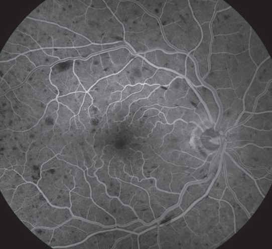
The Central Vein Occlusion Study defined ischemic disease as ten or more disc areas of capillary nonperfusion on fluorescein angiography, and clinicians also use visual acuity, pupil response and the extent of hemorrhage. The line between ischemic and nonischemic disease is not sharp, so repeat the assessment over time.
How Common Is Ischemic CRVO
Most eyes with central retinal vein occlusion are nonischemic at presentation, but a minority are ischemic, and some perfused eyes convert later. The Central Vein Occlusion Study also found that a proportion of perfused eyes became nonperfused within three years, so follow all eyes.
Fundus Explorer Pro
Photograph the retinal findings described here with the phone already in your pocket — 22 D optics and built-in illumination in one handheld unit.
From Choroida — the team behind this siteClinical Signs of Ischemic CRVO
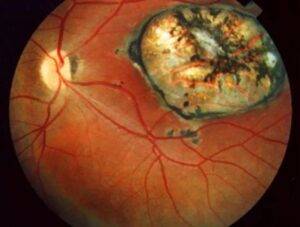
Visual acuity is usually 20/200 or worse, and a relative afferent pupillary defect is common. The retina also shows dense hemorrhages in all quadrants, venous dilation and cotton-wool spots (see central retinal vein occlusion), so a dramatic fundus with poor vision suggests ischemia.
Fluorescein Angiography
Angiography shows large areas of nonperfusion and blocked fluorescence from hemorrhage, and it helps confirm the diagnosis when the signs are mixed. Ultra-widefield imaging also reveals peripheral nonperfusion (see fluorescein angiography), but because the test is not always possible, use the clinical signs too.
Risk of Neovascularization
Hayreh and others described a high rate of neovascularization in ischemic eyes, and iris and angle vessels often appear within the first six months. The risk is higher with extensive nonperfusion and poor acuity, so examine the iris and the angle regularly (see rubeosis iridis).
Monitoring Schedule for Ischemic CRVO
Review monthly for the first six months, and at each visit check the iris and the angle by gonioscopy before dilation. Measure the pressure and examine the fundus too, because angle neovascularization can precede changes on the iris, which makes gonioscopy essential.
Treatment of Neovascularization
Panretinal photocoagulation reduces the stimulus for vessel growth, and the Central Vein Occlusion Study did not support prophylactic treatment but supported prompt treatment when neovascularization appears (see panretinal photocoagulation). Anti-VEGF injections also cause rapid regression of vessels, so combine injections with laser in many eyes.
Neovascular Glaucoma
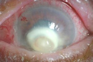
Neovascular glaucoma causes a rapid rise in pressure and severe pain, so treat the pressure, the neovascularization and the pain together (see neovascular glaucoma). Tube surgery or cyclodestruction may also be needed, and because the visual outcome can be poor, early action matters.
Macular Edema in Ischemic CRVO
Macular edema accompanies many eyes, and anti-VEGF drugs reduce the thickness and may improve vision. The improvement is also smaller in ischemic eyes because of macular damage, so set realistic expectations.
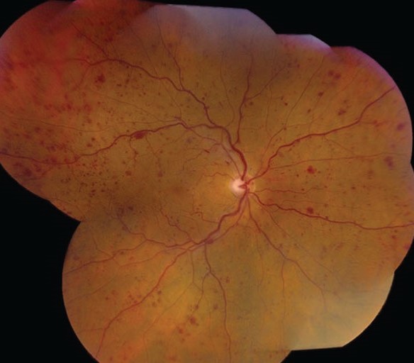
Systemic Evaluation
Check the blood pressure, glucose and lipids, and ask about glaucoma, hyperviscosity and clotting disorders in young patients. Refer for systemic assessment when risk factors are present, and because the other eye can be affected, treat the systemic causes.
Prognosis of Ischemic CRVO
Vision often stays poor in ischemic eyes, although some patients maintain a useful level of vision in the fellow eye. Neovascularization can also be prevented in many eyes with timely treatment.
Counseling the Patient
Explain the risk of pain and vision loss from glaucoma, and explain the schedule of visits and the reason for each. Because adherence matters, give clear written instructions.
Bedside Clues to Ischemia
Check the pupil for a relative afferent defect, look at the retina for dense hemorrhages and multiple cotton-wool spots, and ask about visual acuity. Because bedside signs help when angiography is delayed, use them to decide on urgency.
Role of Optical Coherence Tomography Angiography
OCTA shows the macular capillary dropout without dye, and it can help judge ischemia in cooperative patients. Wide-field systems also extend the view, so it may complement fluorescein.
Anti-VEGF and Retinal Perfusion
Injections do not restore perfusion, but they treat edema and new vessels. Treatment may also mask the need for laser if the eye is not followed, so continue to monitor because ischemia persists.
Systemic Associations
Hypertension, diabetes and hyperlipidemia are common, and glaucoma and hyperviscosity states also contribute. Therefore, send the patient for a medical review.
Follow-Up in the Long Term
Check the fellow eye and the systemic risk factors, and remind patients to return if pain occurs. Because late neovascularization can occur, do not stop follow-up too early.


Document what you see
Two smartphone imaging tools built for everyday clinic use — one for the slit lamp, one for the fundus.
From Choroida — the team behind this siteReferences
- Central Vein Occlusion Study Group. Natural history and clinical management of central retinal vein occlusion. Arch Ophthalmol. 1997;115:486-491.
- Central Vein Occlusion Study Group. A randomized clinical trial of early panretinal photocoagulation for ischemic central vein occlusion: the CVOS Group N report. Ophthalmology. 1995;102:1434-1444.
- Hayreh SS, Klugman MR, Beri M, Kimura AE, Podhajsky P. Differentiation of ischemic from non-ischemic central retinal vein occlusion during the early acute phase. Graefes Arch Clin Exp Ophthalmol. 1990;228:201-217.
- McIntosh RL, Rogers SL, Lim L, et al. Natural history of central retinal vein occlusion: an evidence-based systematic review. Ophthalmology. 2010;117:1113-1123.

Writes and reviews the clinical articles on Educate. Every post is checked against peer-reviewed sources before it goes live.