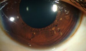
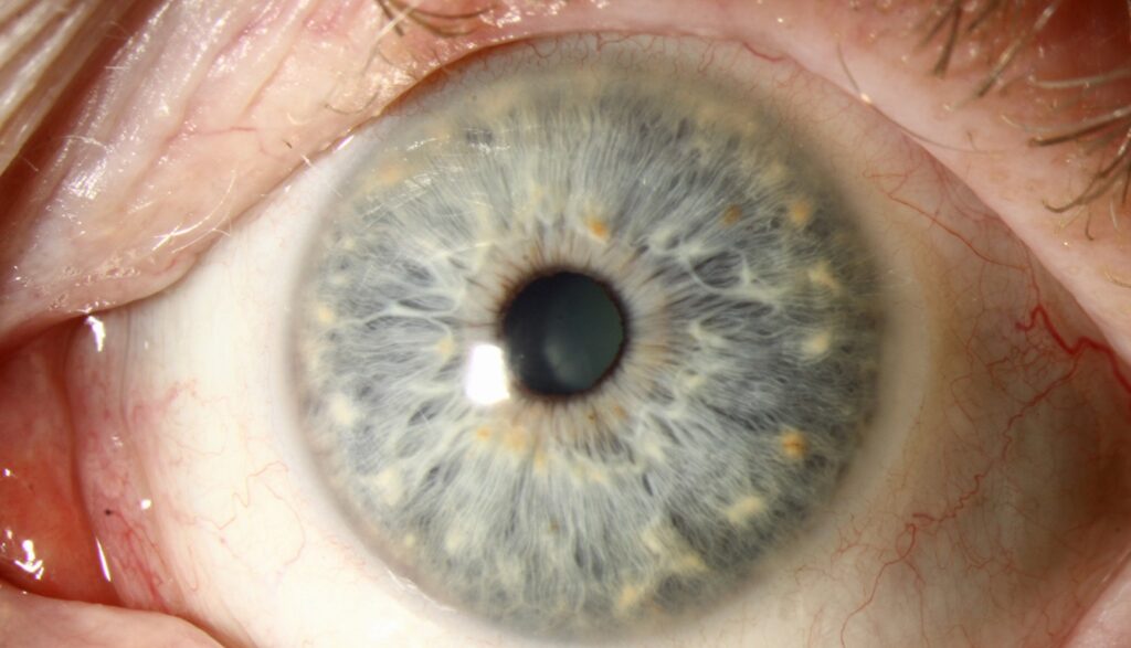
Wolfflin nodules are small, pale elevations in the peripheral iris stroma of otherwise healthy eyes.
Many texts also call them Kruckmann-Wolfflin bodies.
Clinicians usually notice them in light blue or gray irides during a routine slit-lamp examination.
Because they resemble Brushfield spots, they sometimes cause needless concern.
What Wolfflin Nodules Look Like
Each nodule is a tiny gray-white or cream dot on the anterior iris surface.
The dots sit near the iris root, in the far periphery.
They are usually sparse and irregularly spaced.
In addition, the stroma between them looks normal in thickness and texture.
Who Has Wolfflin Nodules
These spots occur in a minority of the general population.
Donaldson found similar peripheral spots in some control eyes while he studied Brushfield spots.
They are far easier to see in lightly pigmented irides.
Therefore, examiners rarely report them in dark brown eyes, where dense stroma hides them.
All-fit Slit-Lamp Adapter
Record and share exactly what you see at the slit lamp. One adapter fits any slit lamp or surgical microscope — and any smartphone.
From Choroida — the team behind this siteHistology in Wolfflin Nodules
Pathologists describe these nodules as focal condensations of normal iris stroma.
Published descriptions show fibrovascular tissue without neoplastic cells.
Brushfield spots share a similar structure.
However, in Down syndrome the surrounding stroma is often hypoplastic, so the spots stand out more.
Symptoms and Visual Function
Wolfflin nodules cause no symptoms.
Vision, pupil reactions and intraocular pressure remain normal.
Patients usually learn about them only when an examiner mentions them.
As a result, the main clinical task is accurate labeling and reassurance.
Slit-Lamp Technique for Wolfflin Nodules
Examine the iris before dilation, because mydriasis bunches the peripheral stroma.
First, use diffuse illumination at low magnification.
Then switch to a narrow oblique beam to judge elevation.
Retroillumination adds little, since the nodules lie in the stroma.
Wolfflin Nodules Versus Brushfield Spots
Brushfield spots show a strong association with Down syndrome (see Brushfield spots in Down syndrome).
They are usually more numerous and form a fairly regular ring in the mid-periphery.
In addition, a zone of stromal hypoplasia often surrounds them.
Wolfflin nodules, by contrast, are fewer, smaller and closer to the iris root.
Wolfflin Nodules Versus Lisch Nodules
Lisch nodules are melanocytic hamartomas seen in neurofibromatosis type 1.
They are tan to brown, dome-shaped and clearly raised.
Their number tends to rise with age, and they can appear anywhere on the iris.
Therefore, color helps first, because Lisch nodules are pigmented while Wolfflin nodules are pale.
Inflammatory Iris Nodules
Koeppe and Busacca nodules signal granulomatous uveitis (see Koeppe and Busacca nodules).
Koeppe nodules sit at the pupillary margin, while Busacca nodules lie on the mid-stroma.
These eyes usually show anterior chamber cells, keratic precipitates or synechiae.
A quiet eye with stable peripheral dots points away from inflammation.
Differential Diagnosis of Wolfflin Nodules
- Brushfield spots: more numerous, mid-peripheral ring, surrounding hypoplasia
- Lisch nodules: brown, dome-shaped, linked to neurofibromatosis type 1
- Freckles: flat, brown spots in the anterior border layer (see iris freckles)
- Nevus: pigmented, may distort the stroma, needs photographic follow-up
- Mammillations: uniform, velvety projections, often on a dark iris
- Granulomatous nodules: fluffy, gray-white, with signs of active uveitis
When to Look Further
A pale iris nodule needs more thought when the history or examination does not fit.
For example, cafe au lait macules or axillary freckling suggest neurofibromatosis.
Developmental delay or typical facial features may prompt a pediatric review.
However, the iris finding alone should never establish a syndrome.
Similarly, any nodule that grows, bleeds or raises pressure needs a full workup.
Documentation of Wolfflin Nodules
Anterior segment photography records the number and position of spots.
Photos also help a later examiner judge stability.
Anterior segment OCT is rarely needed.
Still, it can confirm a superficial stromal lesion when doubt persists.
Counseling Patients
Explain that the spots are a normal variation of iris structure.
They do not affect sight, and they do not become cancer.
Parents sometimes ask whether the spots mean Down syndrome.
A clear, calm explanation of the difference usually ends the worry.
Management and Follow-Up
Wolfflin nodules need no treatment.
Routine eye examinations are enough for typical lesions.
No special imaging schedule is required.
However, document any atypical or pigmented spot and recheck it at the next visit.
Why the Name Matters
Correct terminology avoids labeling a healthy child with a syndromic sign.
A referral letter that says “Brushfield spots” can trigger unnecessary genetic testing.
Therefore, describe the lesion precisely, including location, color, number and surrounding stroma.
Careful wording also protects families from false alarms.
Pitfalls to Avoid
- Do not diagnose Down syndrome from iris spots alone
- Do not call pale peripheral spots Lisch nodules, because Lisch nodules are pigmented
- Avoid judging the iris surface only after dilation
- Do not miss cells or keratic precipitates that point to uveitis
- Never ignore a spot that grows or changes color


Document what you see
Two smartphone imaging tools built for everyday clinic use — one for the slit lamp, one for the fundus.
From Choroida — the team behind this siteReferences
- Donaldson DD. The significance of spotting of the iris in mongoloids: Brushfield’s spots. Arch Ophthalmol. 1961;65:26-31.
- Brushfield T. Mongolism. Br J Child Dis. 1924;21:241-258.
- Lewis RA, Riccardi VM. Von Recklinghausen neurofibromatosis: incidence of iris hamartomata. Ophthalmology. 1981;88:348-354.
- Ragge NK, Falk RE, Cohen WE, Murphree AL. Images of Lisch nodules across the spectrum. Eye. 1993;7:95-101.
- Salmon JF. Kanski’s Clinical Ophthalmology: A Systematic Approach. 9th ed. Elsevier; 2020.
- American Academy of Ophthalmology. Basic and Clinical Science Course, Section 4: Ophthalmic Pathology and Intraocular Tumors. San Francisco: AAO.

Writes and reviews the clinical articles on Educate. Every post is checked against peer-reviewed sources before it goes live.