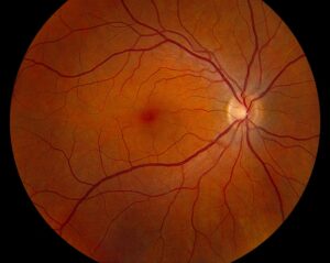
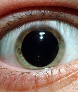
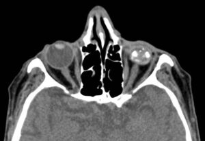
Miosis means a constricted pupil, usually below about 2 mm in ordinary room light.
It may affect one eye or both, and the pattern points toward the cause.
A unilateral small pupil suggests Horner syndrome, uveitis or a local drug.
Bilateral miosis, however, more often reflects systemic drugs, age or neurosyphilis.
How the Pupil Becomes Small
Parasympathetic fibers drive the iris sphincter and constrict the pupil.
In contrast, the dilator muscle depends on an intact three-neuron sympathetic chain.
Miosis therefore follows either sphincter overaction or loss of dilator tone.
Iris inflammation and adhesions can also fix the pupil mechanically.
First Question: Which Pupil Is Abnormal?
With anisocoria, the examiner must first decide which pupil is abnormal.
Lighting answers this quickly.
If the difference grows in darkness, the small pupil fails to dilate.
If it grows in bright light, the large pupil fails to constrict, and the mydriasis workup applies.
All-fit Slit-Lamp Adapter
Record and share exactly what you see at the slit lamp. One adapter fits any slit lamp or surgical microscope — and any smartphone.
From Choroida — the team behind this siteThe Dim-Light Test for Miosis
Measure both pupils in bright room light with a pupil gauge.
Then turn the lights off and watch the pupils with dim oblique light from below.
Next, compare the anisocoria at about 5 seconds and again at 15 seconds.
A larger difference at 5 seconds indicates dilation lag, a classic sign of sympathetic denervation.
Physiological Anisocoria
Simple anisocoria is common in healthy people.
The difference is usually 1 mm or less.
Also, it stays roughly constant in light and dark, and it shows no dilation lag.
Old photographs help, because the asymmetry may vary over time or switch sides.
Horner Syndrome Miosis
Horner syndrome combines mild ptosis, miosis and sometimes facial anhidrosis.
The ptosis is subtle because only Muller muscle loses tone.
In addition, the lower lid may sit slightly higher, giving apparent enophthalmos.
In congenital cases, the affected iris can look lighter.
Confirming Horner Syndrome
Apraclonidine 0.5% or 1% dilates a denervated pupil through alpha-1 supersensitivity.
As a result, the anisocoria reverses, and the ptosis often improves.
Cocaine 10% is the older test, and the Horner pupil dilates poorly after it.
However, apraclonidine may give a false negative in very recent lesions, because supersensitivity takes time to develop.
Urgent Horner Presentations
Acute painful Horner syndrome warrants same-day vascular imaging for carotid dissection.
Neck pain, headache or recent neck trauma raises suspicion.
Similarly, a new Horner pupil in a smoker may reflect an apical lung tumor.
Miosis in Anterior Uveitis
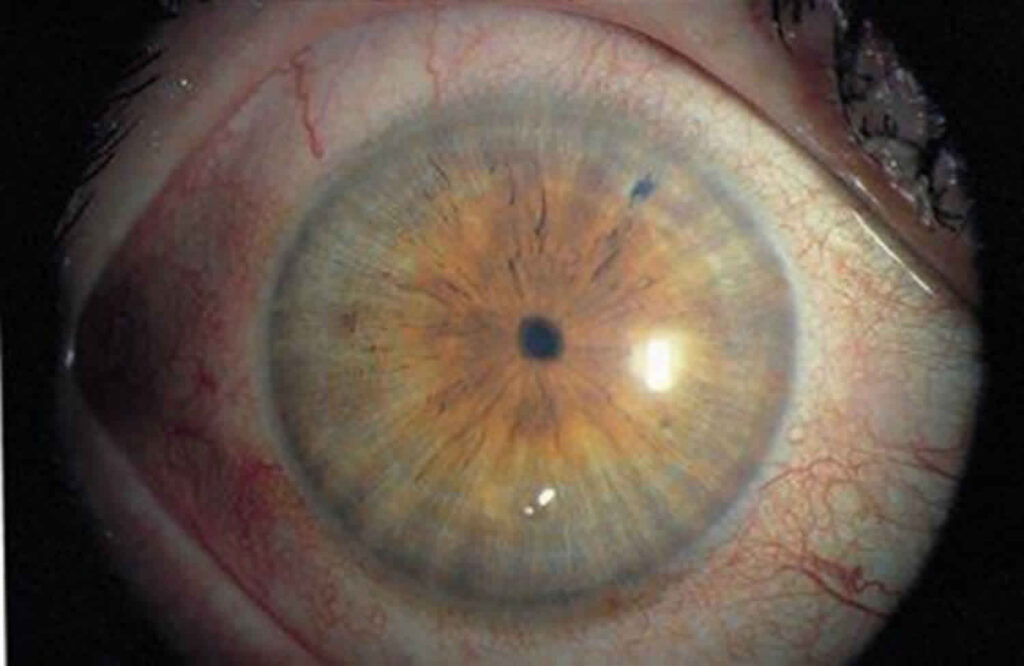
Inflammation irritates the iris sphincter, so the pupil constricts and reacts sluggishly.
The patient reports pain, photophobia and redness, often with ciliary flush.
At the slit lamp, look for cells, flare and keratic precipitates (see acute anterior uveitis).
Posterior synechiae then create an irregular pupil that dilates unevenly (see posterior synechiae).
Treating Miosis in Uveitis
Cycloplegic drops relax the sphincter and ease ciliary spasm pain.
They also help break fresh synechiae and prevent new ones.
However, they do not control the inflammation, so topical steroids stay the main treatment.
Tell the patient that blurred near vision and glare are expected while the pupil is dilated.
Topical Drug-Induced Miosis
Pilocarpine is the classic cause, from glaucoma treatment or presbyopia drops.
Brimonidine and other alpha-2 agonists can produce mild miosis, especially in dim light.
Ask about every drop, including products borrowed from family members.
Miosis From Systemic Drugs
Opioids cause bilateral pinpoint pupils that still react weakly to light.
Organophosphate insecticides produce severe miosis with other cholinergic signs.
Therefore, a drowsy patient with tiny pupils needs a medication and exposure history.
Pinpoint pupils with reduced consciousness also require urgent general medical assessment.
Argyll Robertson Pupils
These are small, irregular pupils, usually bilateral and often asymmetric.
They react poorly to light yet constrict briskly to a near target.
This light-near dissociation should prompt serologic testing for syphilis (see Argyll Robertson pupil).
They also dilate poorly with mydriatic drops.
An Exam Sequence for Miosis
- Measure pupils in bright light and in darkness.
- Check for dilation lag at 5 and 15 seconds.
- Test light and near reactions in each eye.
- Look for upper lid ptosis and lower lid elevation.
- Examine the iris, anterior chamber and pupil margin at the slit lamp.
- Review drops, systemic drugs and old photographs.
- Confirm Horner syndrome pharmacologically when suspected.
Pitfalls to Avoid
- Do not test pupils only in bright light
- Never overlook posterior synechiae when an irregular small pupil dilates poorly
- Do not attribute acute painful Horner syndrome to migraine without imaging
- Ask about presbyopia drops before ordering any workup


Document what you see
Two smartphone imaging tools built for everyday clinic use — one for the slit lamp, one for the fundus.
From Choroida — the team behind this siteReferences
- Thompson HS, Pilley SF. Unequal pupils. A flow chart for sorting out the anisocorias. Surv Ophthalmol. 1976;21:45-48.
- Lam BL, Thompson HS, Corbett JJ. The prevalence of simple anisocoria. Am J Ophthalmol. 1987;104:69-73.
- Kardon RH, Denison CE, Brown CK, Thompson HS. Critical evaluation of the cocaine test in the diagnosis of Horner’s syndrome. Arch Ophthalmol. 1990;108:384-387.
- Koc F, Kavuncu S, Kansu T, Acaroglu G, Firat E. The sensitivity and specificity of 0.5% apraclonidine in the diagnosis of oculosympathetic paresis. Br J Ophthalmol. 2005;89:1442-1444.
- Jabs DA, Nussenblatt RB, Rosenbaum JT; SUN Working Group. Standardization of uveitis nomenclature for reporting clinical data. Am J Ophthalmol. 2005;140:509-516.

Writes and reviews the clinical articles on Educate. Every post is checked against peer-reviewed sources before it goes live.