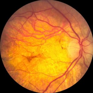
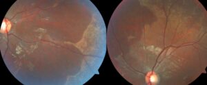
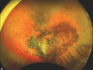
Optic neuritis recovery usually begins within the first few weeks, and most patients regain useful vision.
However, the timeline varies, and normal acuity does not always mean normal visual function.
Therefore, clinicians should know the expected course and recognize when a patient departs from it.
Typical Optic Neuritis Recovery Timeline
Vision commonly worsens over several days, and it may decline for up to two weeks.
Improvement then usually starts within about three weeks of onset.
Most of the gain occurs during the first few months.
However, slower improvement can continue for up to a year.
Long-Term Vision in the ONTT
The Optic Neuritis Treatment Trial provides the best long-term prognosis data (see ONTT results).
At 15 years, 72% of affected eyes saw 20/20 or better.
In addition, about 3% saw 20/200 or worse.
However, these figures come from a largely typical demyelinating cohort, so they may not apply to atypical disease.
Fundus Explorer Pro
Photograph the retinal findings described here with the phone already in your pocket — 22 D optics and built-in illumination in one handheld unit.
From Choroida — the team behind this sitePain During Optic Neuritis Recovery
Pain on eye movement often precedes the visual loss.
It usually settles within days, often before vision improves.
Therefore, pain that persists beyond the expected period deserves a second look.
Residual Symptoms After Optic Neuritis Recovery
Patients with good acuity may still report that the affected eye looks different.
These complaints reflect permanent axonal and myelin injury, and Snellen testing can miss them.
Common residual symptoms include:
- Reduced color saturation, especially for red
- Poor contrast sensitivity in dim light
- Reduced depth or motion perception
- Patchy visual field defects
Uhthoff Phenomenon
Some patients notice transient blur during exercise, fever or a hot shower.
Heat temporarily impairs conduction along demyelinated axons, and vision returns as the body cools.
In contrast, new loss that persists for days requires examination.
Examination Signs During Optic Neuritis Recovery
A relative afferent pupillary defect can persist after acuity returns.
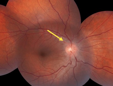
Temporal or diffuse disc pallor often appears over the following weeks and months.
Color vision and contrast testing also reveal deficits that acuity misses.
OCT in Optic Neuritis Recovery
Early disc swelling can mask axonal loss on peripapillary retinal nerve fiber layer scans.
As edema resolves, thinning becomes apparent over the next several months.
Ganglion cell layer analysis often reveals macular thinning earlier.
Do Steroids Change Optic Neuritis Recovery?
In the ONTT, intravenous methylprednisolone followed by oral prednisone sped visual recovery.
However, it did not improve long-term acuity in typical disease.
Standard-dose oral prednisone alone did not improve vision, and the trial linked it to more recurrences.
High-dose oral regimens also showed noninferior outcomes in a bioequivalence trial.
Risks include insomnia, mood change and hyperglycemia, so discuss the modest benefit before treatment.
Red Flags for Reassessment
Atypical features should prompt review of the diagnosis and new investigations.
Warning signs include:
- No improvement after about three to four weeks
- Progressive loss beyond two weeks
- Painless onset, particularly with age over 50
- Bilateral simultaneous involvement
- No light perception at presentation
- Marked disc edema, hemorrhages or macular exudates
- Relapse when steroids taper
Antibody Testing When Optic Neuritis Recovery Stalls
Severe or poorly recovering neuritis should raise suspicion for aquaporin-4 antibody disease.
These patients often need urgent escalation, and clinicians may consider plasma exchange.
MOG antibody disease often shows disc swelling and good steroid response, but relapses can follow tapering (see MOG-IgG optic neuritis).
Mimics to Exclude
A failed recovery often means the first diagnosis was wrong.
For example, sudden painless altitudinal loss in an older patient suggests NAION.
Sequential painless central loss in a young adult suggests Leber hereditary optic neuropathy.
In addition, consider compressive lesions, sarcoidosis, syphilis and toxic or nutritional neuropathy.
MS Risk After Optic Neuritis Recovery
Good visual recovery does not lower concern about future multiple sclerosis.
In the ONTT, 15-year risk reached 50% overall.
Risk was 25% with no baseline brain lesions, and 72% with one or more lesions.
Therefore, review the MRI and arrange neurology input early.
Counseling and Follow-Up
Tell patients that vision often starts improving within weeks, although individual courses vary.
Next, explain that color and contrast may stay subtly reduced after acuity recovers.
Also, ask them to report new loss or neurological symptoms promptly.
Then repeat acuity, color vision, pupils, fields and OCT at intervals matched to the course.
Finally, record the stable result as the baseline for judging any recurrence.
Pitfalls to Avoid
- Do not call recovery complete based on acuity alone
- Never treat Uhthoff symptoms as an automatic relapse
- Do not read early normal OCT as proof of axonal preservation
- Avoid standard-dose oral prednisone alone for typical disease


Document what you see
Two smartphone imaging tools built for everyday clinic use — one for the slit lamp, one for the fundus.
From Choroida — the team behind this siteReferences
- Beck RW, Cleary PA, Anderson MM, et al. A randomized, controlled trial of corticosteroids in the treatment of acute optic neuritis. N Engl J Med. 1992;326:581-588.
- Optic Neuritis Study Group. Visual function 15 years after optic neuritis: a final follow-up report from the Optic Neuritis Treatment Trial. Ophthalmology. 2008;115:1079-1082.
- Optic Neuritis Study Group. Multiple sclerosis risk after optic neuritis: final Optic Neuritis Treatment Trial follow-up. Arch Neurol. 2008;65:727-732.
- Morrow SA, Fraser JA, Day C, et al. Effect of treating acute optic neuritis with bioequivalent oral vs intravenous corticosteroids. JAMA Neurol. 2018;75:690-696.
- Petzold A, Fraser CL, Abegg M, et al. Diagnosis and classification of optic neuritis. Lancet Neurol. 2022;21:1120-1134.

Writes and reviews the clinical articles on Educate. Every post is checked against peer-reviewed sources before it goes live.