Preoperative anti-VEGF injection before diabetic vitrectomy shrinks neovascular tissue and lowers intraoperative bleeding.
Surgeons use it in eyes with active fibrovascular proliferation, vitreous hemorrhage, or tractional detachment.
The timing matters, since a long delay can worsen traction.
Why Preoperative Anti-VEGF Helps
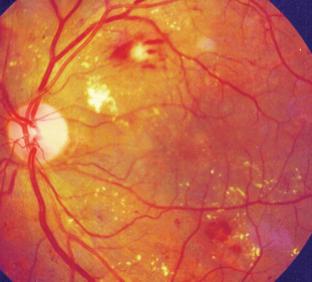
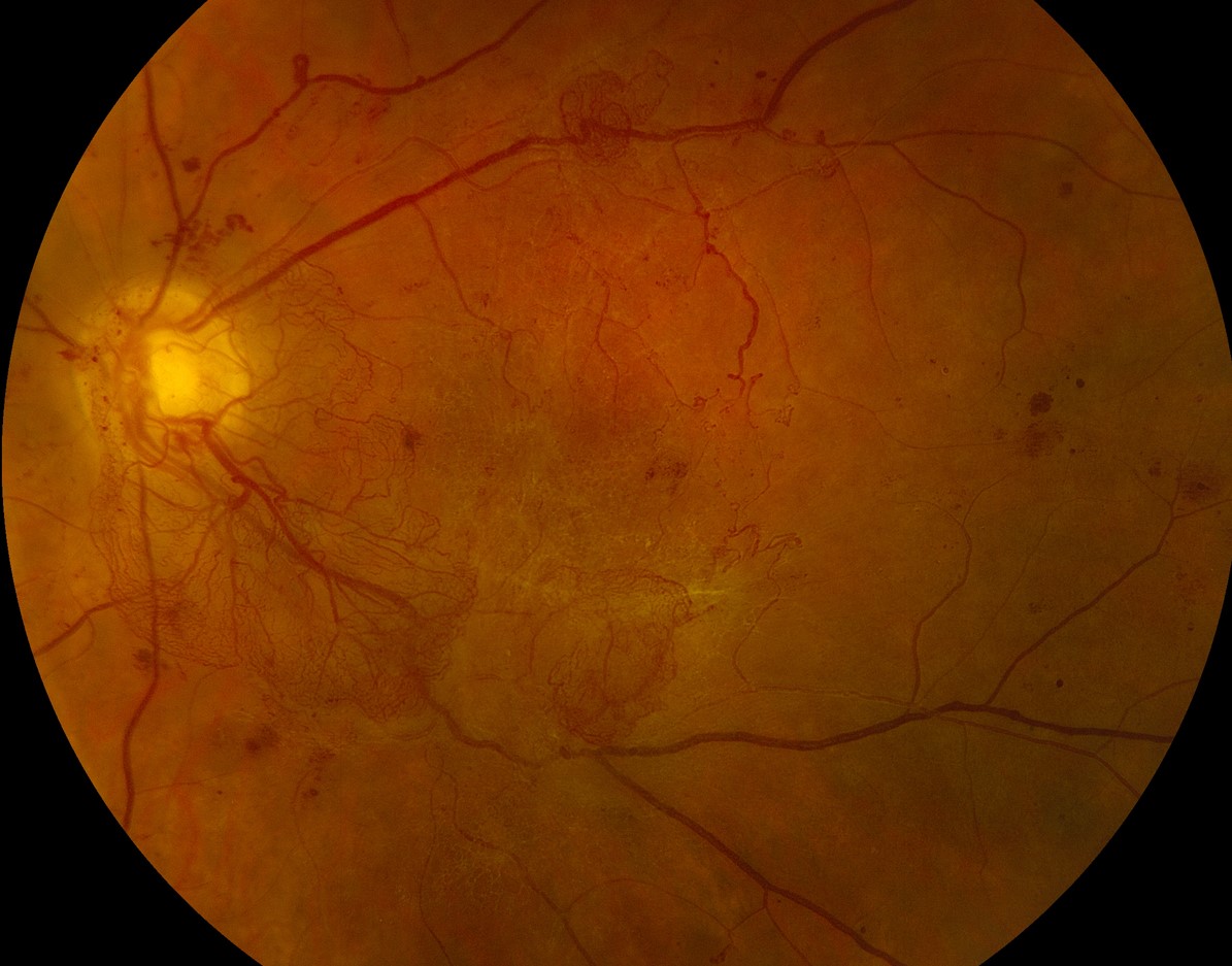
Fibrovascular membranes in proliferative diabetic retinopathy contain abundant new vessels that bleed easily.
Anti-VEGF drugs cause these vessels to regress within days, so dissection becomes easier.
Less bleeding gives a clearer view, shorter operating time, and less need for diathermy (see proliferative diabetic retinopathy).
Timing of Preoperative Anti-VEGF
Most surgeons inject three to seven days before surgery.
The effect on vessels peaks within the first week and fades afterward.
A longer interval increases the risk of the crunch phenomenon, in which regressing vessels cause the fibrous tissue to contract and the retina to detach or tear.
Arevalo and colleagues described this risk in patients with severe proliferative disease.
Fundus Explorer Pro
Photograph the retinal findings described here with the phone already in your pocket — 22 D optics and built-in illumination in one handheld unit.
From Choroida — the team behind this siteEvidence for Preoperative Anti-VEGF
Randomized trials and meta-analyses show lower rates of intraoperative bleeding and early postoperative vitreous hemorrhage with pretreatment.
Operating times and the need for silicone oil tend to decrease, although visual outcomes differ little between groups.
A Cochrane review found that anti-VEGF lowers early postoperative hemorrhage after vitrectomy for proliferative disease.
Trial quality varies, so the size of the benefit remains uncertain.
Which Eyes Benefit Most From Preoperative Anti-VEGF
- Dense, vascular fibrovascular plaques
- Active neovascularization with recent bleeding
- Severe tractional detachment before surgery
Eyes with fibrotic membranes and little active neovascularization gain less (see tractional retinal detachment).
Risks of Preoperative Anti-VEGF
The crunch phenomenon is the main concern, and it needs careful timing.
Inject only as many days before surgery as the schedule permits.
Examine the eye before injection to rule out significant traction on the macula, since progression may follow.
Also review the patient’s systemic risk, such as recent stroke, because anti-VEGF drugs enter the circulation (see anti-VEGF therapy).
Intraoperative Anti-VEGF
Some surgeons inject at the end of surgery instead, to reduce the risk of postoperative bleeding.
This approach avoids the crunch phenomenon, though it gives no help during dissection.
Others combine both approaches in complex eyes.
An Example Case
A 44-year-old man with type 1 diabetes has a dense vitreous hemorrhage with fibrovascular plaques and a tractional detachment.
Surgery is scheduled for a Friday, and he receives an intravitreal injection of bevacizumab on the preceding Monday.
At surgery, the plaques separate easily, bleeding is minimal, and the retina flattens after peeling.
The surgeon completes laser and leaves gas, and the retina remains attached at three months.
Questions Patients Ask
Patients ask why they need an injection before surgery, and the answer is that it makes the operation safer and easier.
They also ask whether the injection is painful, and most feel only brief pressure.
Because surgery should follow soon after, explain that delays may increase the risk of traction.
Pitfalls to Avoid
- Injecting more than a week before surgery in eyes with significant traction
- Using the injection as a substitute for panretinal laser when surgery is not planned
- Ignoring recent stroke or heart disease before the injection
- Forgetting to examine the macula for progression before surgery
Looking Ahead
New anti-VEGF agents may offer more durable regression, and trials will test them in this setting.
Researchers also study whether pretreatment improves long-term vision, beyond surgical ease.
Until then, use preoperative injection selectively in eyes with active vascular proliferation.
Practical Points
Schedule the injection and the operating room date together, so the interval stays short.
Then examine the eye on the day of surgery to check the status of traction.
Also record the injection date and drug in the operative note.
Choice of Drug
Bevacizumab, ranibizumab, and aflibercept have all been used before vitrectomy.
Bevacizumab costs less and has the most reports, while the others show similar effects in trials.
Therefore, choose by cost, availability, and the surgeon’s experience.
Also follow local rules for off-label use and document consent.
Combined Treatment
Many surgeons also give panretinal laser at the time of surgery to lower the risk of new vessels.
Then they use the injection to ease the dissection and reduce bleeding.
Together, these steps lower the chance of repeat surgery for hemorrhage.
For the latest studies, see the PubMed results for preoperative anti-VEGF.


Document what you see
Two smartphone imaging tools built for everyday clinic use — one for the slit lamp, one for the fundus.
From Choroida — the team behind this siteReferences
- Arevalo JF, Maia M, Flynn HW Jr, et al. Tractional retinal detachment following intravitreal bevacizumab (Avastin) in patients with severe proliferative diabetic retinopathy. Br J Ophthalmol. 2008;92:213-216.
- Zhao XY, Xia S, Chen YX. Antivascular endothelial growth factor agents pretreatment before vitrectomy for complicated proliferative diabetic retinopathy: a meta-analysis of randomised controlled trials. Br J Ophthalmol. 2018;102:1077-1085.
- Smith JM, Steel DH. Anti-vascular endothelial growth factor for prevention of postoperative vitreous cavity haemorrhage after vitrectomy for proliferative diabetic retinopathy. Cochrane Database Syst Rev. 2015;(8):CD008214.
- American Academy of Ophthalmology. Basic and Clinical Science Course, Section 12: Retina and Vitreous. AAO; current edition.

Writes and reviews the clinical articles on Educate. Every post is checked against peer-reviewed sources before it goes live.