Chalcosis is the toxic reaction of ocular tissues to copper from an intraocular or intralenticular foreign body.
Pure copper causes a violent inflammatory reaction, while alloys such as brass and bronze cause a slower deposition.
Therefore, you must decide quickly whether a copper-rich fragment sits in the eye.
Why Copper Is Toxic
Copper has an affinity for basement membranes, such as Descemet membrane, the lens capsule and the inner limiting membrane.
It deposits there and drives oxidative damage.
Because copper ions are soluble in ocular fluids, they spread through the globe.
Acute and Chronic Chalcosis
Fragments with a high copper content, above about 85 percent, provoke an acute purulent reaction that can mimic endophthalmitis.
Alloys with a lower copper content cause a chronic, smouldering course.
Also, the reaction depends on the size, the site and the time the fragment stays in the eye.
All-fit Slit-Lamp Adapter
Record and share exactly what you see at the slit lamp. One adapter fits any slit lamp or surgical microscope — and any smartphone.
From Choroida — the team behind this siteAnterior Segment Signs of Chalcosis
Copper deposits in Descemet membrane and form a greenish-brown ring that looks like a Kayser-Fleischer ring (see Kayser-Fleischer ring).
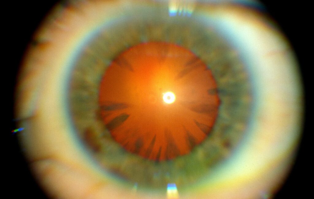
The lens may develop a sunflower cataract with petal-like spokes (see sunflower cataract).
The iris can turn a greenish color, and the pupil can react poorly.
Posterior Segment Signs of Chalcosis
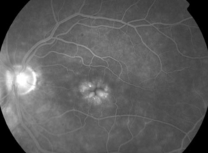
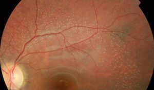
Vitreous opacities and refractile golden deposits appear at the inner limiting membrane.
Also, retinal vessels may show a metallic sheen.
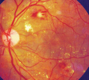
Later, macular edema, retinal degeneration and optic atrophy follow.
Therefore, retinal function may be lost if the fragment stays for long.
Diagnosing Chalcosis
Take a history of hammering metal on metal, grinding, explosions or firearm injury.
Computed tomography is the imaging method of choice for a suspected metallic foreign body.
Also, B-scan ultrasound and ultrasound biomicroscopy help for fragments near the angle.
Avoid magnetic resonance imaging until you have excluded a magnetic fragment.
Management of Chalcosis
Remove a copper-rich foreign body promptly.
Vitrectomy with forceps is the usual approach, since copper is not magnetic and a magnet will not extract it.
Also, give tetanus prophylaxis and consider antibiotic cover for the risk of infection.
Repair the globe and manage the lens as needed (see open globe injury).
Prognosis
Early removal gives the best outcome, but existing deposits may persist.
Visual results depend on the copper content, the time to removal and retinal damage at presentation.
Because the trauma itself can cause damage, tell the patient that vision may stay reduced.
Chalcosis Versus Siderosis
Iron fragments cause siderosis, which brings brown deposits, a dilated pupil and retinal degeneration (see siderosis bulbi).
A magnet attracts iron, and it does not attract copper.
Therefore, the composition of the fragment guides both the diagnosis and the surgical method.
Mechanisms of Retinal Toxicity
Copper ions bind to proteins and damage photoreceptors and the retinal pigment epithelium.
Also, they trigger oxidative stress and inflammation.
Therefore, retinal function can fall even when the media look clear.
Electrophysiology
The electroretinogram can show a reduced b-wave in toxic retinopathy from metal fragments.
Also, serial recordings help follow function after removal.
Because ERG changes may precede visible damage, use it when the fragment stays in place for a while (see electroretinography).
Injury Patterns
Metal on metal injuries often produce high-speed fragments that enter without pain.
Also, patients may not recall the event.
Therefore, ask carefully about hammering, grinding and fireworks, and examine the entry wound.
Risk of Endophthalmitis
Intraocular foreign bodies carry a risk of infection.
Also, wounds from workshop injuries may be contaminated.
Therefore, give antibiotics in line with local protocols for open globe injury.
Follow-Up After Removal
Check pressure, lens clarity and retinal function over months.
Also, monitor for proliferative vitreoretinopathy, since trauma and vitrectomy raise the risk (see proliferative vitreoretinopathy).
Because late complications occur, plan long-term review.
Prevention
Encourage eye protection in workshops and when using hammers on metal.
Also, remind patients that safety glasses with side shields protect against flying fragments.
Role of Imaging Beyond Computed Tomography
Ultrasound biomicroscopy images small fragments in the angle or the ciliary body.
Also, B-scan shows fragments in the vitreous and retinal detachment.
Because small fragments can hide in the wall of the globe, combine imaging methods.
Counselling About Visual Outcomes
Explain that the toxic effect of copper can continue even after removal.
Also, tell the patient that cataract and glaucoma can appear years later.
Therefore, regular follow-up matters, and early reporting of new symptoms helps.
Practical Points for the Emergency Visit
Ask whether the injury involved metal on metal and whether the patient wore eye protection.
Examine the entry site, the pupil, the lens and the fundus when the view permits.
Also, shield the eye, avoid pressure, and arrange imaging without delay.
Because a small fragment can cause serious toxicity, keep a low threshold for scanning.
Then plan removal in a vitreoretinal service as soon as the patient is stable.
For the latest studies, see the PubMed results for chalcosis.


Document what you see
Two smartphone imaging tools built for everyday clinic use — one for the slit lamp, one for the fundus.
From Choroida — the team behind this siteReferences
- Rosenthal AR, Appleton B, Hopkins JL. Intraocular copper foreign bodies. Am J Ophthalmol. 1974;78:671-678.
- Rao NA, Tso MOM, Rosenthal AR. Chalcosis in the human eye: a clinicopathologic study. Arch Ophthalmol. 1976;94:1379-1384.
- Hope-Ross M, Mahon GJ, Johnston PB. Ocular siderosis. Eye. 1993;7:419-425.
- Loporchio D, Mukkamala L, Gorukanti K, Zarbin M, Langer P, Bhagat N. Intraocular foreign bodies: a review. Surv Ophthalmol. 2016;61:582-596.

Writes and reviews the clinical articles on Educate. Every post is checked against peer-reviewed sources before it goes live.