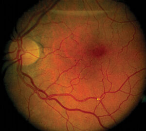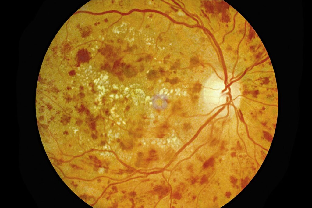
Retinopathy in type 1 diabetes follows a more predictable course than in type 2 diabetes, because the start of the disease is usually known.
The retina is rarely affected in the first years and before puberty, but the risk climbs steadily with duration and with poor control.
Screening guidelines are also built on this timeline.
How Retinopathy in Type 1 Diabetes Behaves
In type 1 diabetes, the onset is often in childhood or adolescence, and the date of diagnosis is well defined.
After five years, a growing number of patients show early retinal changes. After 15 to 20 years, most have some retinopathy, with a significant minority developing proliferative disease if control is poor.
Prepubertal duration seems to count less toward the risk than the years after puberty, which is the basis for starting screening around puberty.
Severe vision-threatening retinopathy is uncommon before the age of 15, but it can occur in those with poor control.
Risk Factors of Retinopathy in Type 1 Diabetes
- Long duration of diabetes
- High HbA1c and glycemic variability, the strongest modifiable factors
- Puberty, with hormonal and growth-related effects on the microvasculature
- Hypertension and dyslipidemia
- Nephropathy and microalbuminuria (see diabetic retinopathy and kidney disease)
- Pregnancy, which can accelerate retinopathy (see diabetic retinopathy in pregnancy)
- Smoking in older patients
- Genetic susceptibility
A rapid improvement in control can cause early worsening in some patients (see early worsening of diabetic retinopathy).
Fundus Explorer Pro
Photograph the retinal findings described here with the phone already in your pocket — 22 D optics and built-in illumination in one handheld unit.
From Choroida — the team behind this siteRetinopathy in Type 1 Diabetes: When to Start Screening
Guidelines from diabetes and ophthalmology bodies recommend the following.
- Begin dilated examinations or retinal photography at about 11 years of age or at puberty, whichever comes first, once the child has had diabetes for two to five years, according to the guideline in use
- Examine adolescents and adults within five years of diagnosis
- Repeat yearly, with longer intervals possible if the retinas are normal and control is good, and shorter intervals if retinopathy is present
- Arrange an examination before pregnancy and in the first trimester for women with type 1 diabetes
Many programs use retinal photography with grading by trained readers, with referral to a specialist for referable disease (see AI diabetic retinopathy screening).
What DCCT and EDIC Showed
The Diabetes Control and Complications Trial randomized patients with type 1 diabetes to intensive or conventional insulin therapy.
Also, intensive therapy reduced the development of retinopathy by about 76 percent in those without it at baseline. It also slowed progression in those with early disease, by about 54 percent (see the references).
The follow-up study, EDIC, showed that the benefit persisted for many years after the trial, even when HbA1c converged, a phenomenon called metabolic memory or the legacy effect.
This finding underlines the value of good control early in the disease.
Features in Young Patients
The retinopathy seen in children and teenagers is usually mild, with microaneurysms and scattered hemorrhages.
Macular edema and proliferative disease are rare in the first decade, but they do occur, and ophthalmic care is needed once they appear.
In addition, the fundus can be difficult to photograph in small children, and screening methods should be adapted to the age.
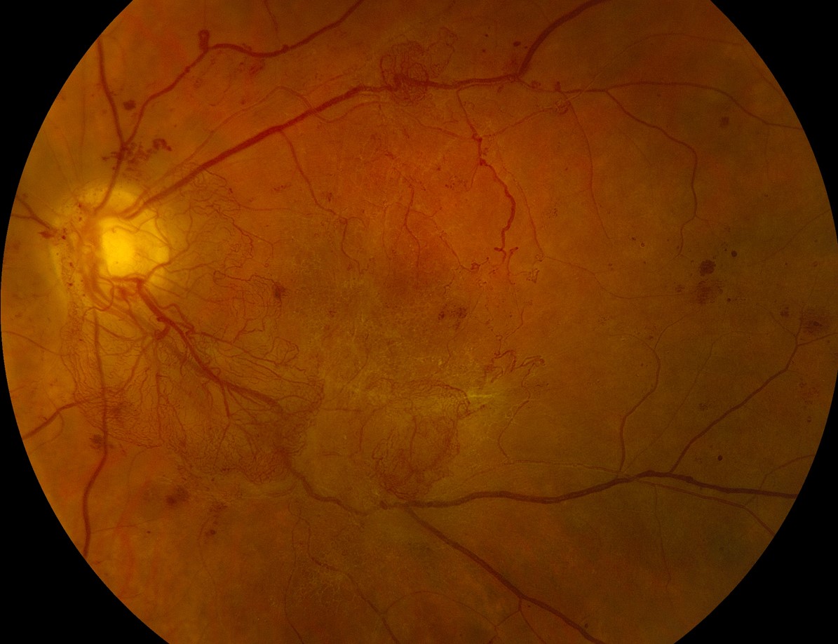
Management for Retinopathy in Type 1 Diabetes
- Systemic control: glycemic control as close to target as safely possible, blood pressure control, and lipid management
- Ophthalmic treatment: the same as for adults, with laser, injections, or surgery for proliferative disease and macular edema (see diabetic retinopathy)
- Care of transitions: from pediatric to adult services, when follow-up often lapses
- Education: young patients benefit from clear explanations about the link between control and eye health
Adolescents often miss appointments, so reminders and a trusting relationship help to keep them in care.
Counselling
Explain to the family that the eye is often normal in the first years, that examinations are a way to catch changes early, and that good control protects the eyes.
Provide advice about driving, sports, and career choices as appropriate, and help the young person to plan for the long-term.
Transition and Adherence
Young people moving from pediatric to adult care often miss eye examinations for several years.
Some return with advanced disease.
Clinics can reduce this by sending reminders, by making appointments before the transfer, and by explaining why retinal screening matters even when vision feels normal.
Involving the family and using digital reminders may help.
Special Situations
Teenagers with poor control and early retinopathy need an intensive support plan, with attention to mental health. Eating disorders and depression are common in this group and can affect control.
Rapid improvement in control should also be accompanied by a retinal check, as discussed elsewhere.


Document what you see
Two smartphone imaging tools built for everyday clinic use — one for the slit lamp, one for the fundus.
From Choroida — the team behind this siteReferences
- Diabetes Control and Complications Trial Research Group. The effect of intensive treatment of diabetes on the development and progression of long-term complications in insulin-dependent diabetes mellitus. N Engl J Med. 1993;329:977-986.
- Nathan DM, Cleary PA, Backlund JY, et al. Intensive diabetes treatment and cardiovascular disease in patients with type 1 diabetes. N Engl J Med. 2005;353:2643-2653.
- Cheung N, Mitchell P, Wong TY. Diabetic retinopathy. Lancet. 2010;376:124-136.
- American Academy of Ophthalmology. Basic and Clinical Science Course, Section 12: Retina and Vitreous. AAO; current edition.

Writes and reviews the clinical articles on Educate. Every post is checked against peer-reviewed sources before it goes live.