
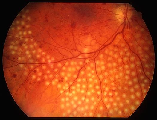
Diabetic retinopathy is a common complication of diabetes and is a major cause of vision impairment and blindness.

It is a condition caused by diabetes that affects the retina. Blood vessels in the retina are damaged and become leaky or blocked.

Abnormal blood vessels can grow from the retina, which can bleed or cause scarring of the retina and result in permanent vision impairment or blindness.
Vision impairment most commonly occurs due to thickening in the central part of the retina (diabetic macular edema), which can lead to irreversible vision impairment.
Several tests are used for diabetic retinopathy screening.
Test performance will need to be considered against other factors, such as cost and ease of use.
The sensitivity of different tests can vary according to who does the examination and how well they are trained, which is important for tests such as direct ophthalmoscopy.
Direct ophthalmoscopy for Diabetic retinopathy

Advantages:
• Mobile
• Relatively inexpensive
• Does not require any special facilities to use
Disadvantages:
• Requires pupil dilation
• Only a small field of the retina can be examined
• Low sensitivity: even with a trained practitioner, small microvascular abnormalities may be difficult to detect
• Less effective than slit-lamp biomicroscopy through dilated pupils
• No ability to audit retrospectively
Indirect ophthalmoscopy for Diabetic retinopathy
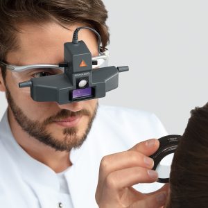
Advantages:
• Mobile
• Large fields of the retina can be examined
• Relatively inexpensive
Disadvantages:
• Requires pupil dilation
• Even with a trained practitioner, small microvascular abnormalities will be difficult to detect
• Less effective than slit-lamp biomicroscopy through dilated pupils
• No ability to audit retrospectively
Slit lamp biomicroscopy for Diabetic retinopathy
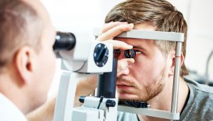
Advantages:
• Large field of the retina can be examined
• Gold standard for training health professionals in retinal examination
• Mobile table-top versions are available
Disadvantages:
• Relatively expensive
• Most types are desk-based
• Requires pupil dilation
• Requires special lenses
• Usually unable to retrospectively audit results, although it is possible to photograph findings
Optical coherence tomography (OCT) for Diabetic retinopathy
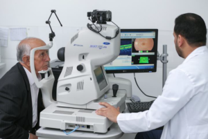
Advantages:
• One of the best ways to assess macular edema (retinal thickening and intraretinal edema)
Disadvantages:
• Needs to be used alongside other screening tests such as slit-lamp or retinal photography to detect diabetic retinopathy
• Relatively expensive
After all these previous methods, We in Choroida decided to innovate a tool that combines all the advantages of the previous devices and avoids their disadvantages.
And we present to you :![]()
Mobile funduscopy ( Choroida fundus explorer ) press here
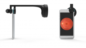
⦁ Simple, easy & affordable
⦁ Suitable for all mobile phones
⦁ 3D Printed
⦁ Portable
⦁ Lightweight 60gm
⦁ Compatible with many fundus lenses (Volk 20D – Volk 28D – Nikon 20D – Occular 20D – Opticlear 20D)
⦁ Depend on mobile led flashlight
⦁ Very Cheap
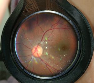
⦁ capture images and record a video
with Fundus Explorer you can record a video of the lesions found; size, color, shape, or even count of hard exudates or dots and blots !!
you can connect the phone to a bigger screen and show the signs live in a conference/ meeting.
you can document a patient’s full exam recorded in a video and upload it online to hospital records.
It makes it so easy that any other professional can continue the treatment plan of Diabetic retinopathy after you and have all data about the patient.
Examine patients at their homes or other departments at the hospital and know about more disease such as Bietti Crystalline Dystrophy, Valsalva Retinopathy,Commotio Retinae.

