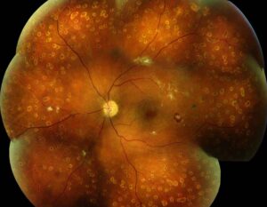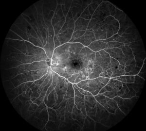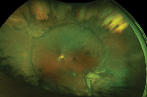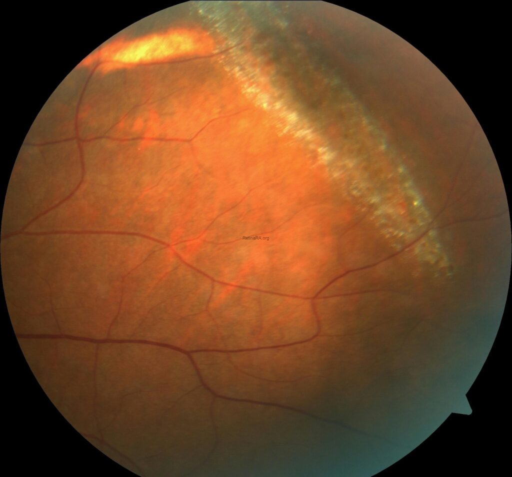
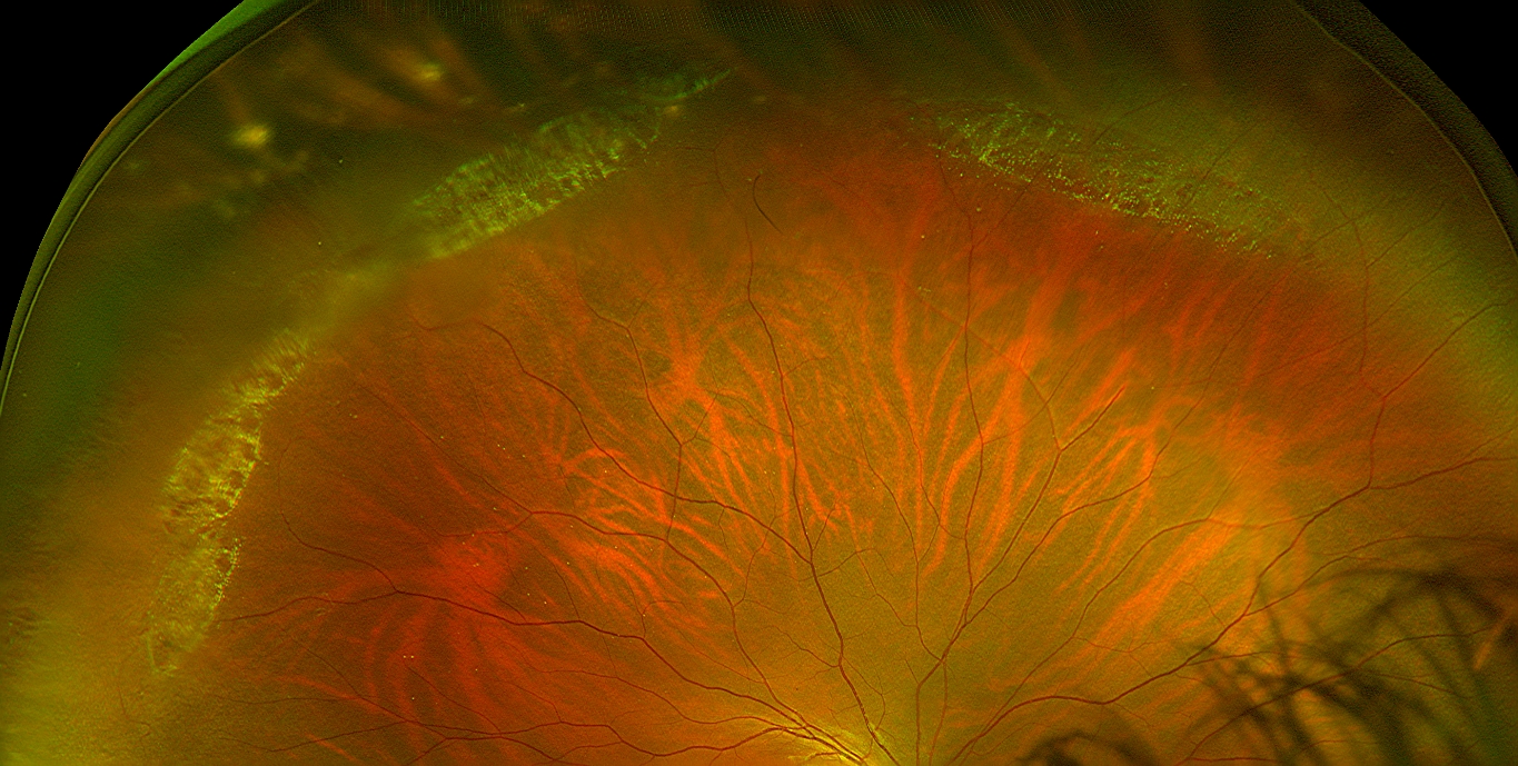
Snail track degeneration is a peripheral retinal change in which a band of tiny, glistening, frost-like dots lies in the retina like the track of a snail.
It is also closely related to lattice degeneration, and doctors often find it together with it.
Most patients have no symptoms, and the finding is also noted at a dilated examination.
What Is Snail Track Degeneration?
The lesion is a sharply demarcated band of retinal thinning, with a silvery white, glistening appearance caused by small dots of reflective material.
It lies parallel to the ora serrata, usually in the superotemporal or inferotemporal periphery, and it may also be circumferential.
Unlike classic lattice, it typically lacks the branching white lines and the pigment, though the two can coexist in the same eye.
Histologic studies have shown retinal thinning, glial changes, and vitreous liquefaction over the lesion, like lattice (see lattice degeneration).
Who Gets It?
- Myopes, in whom peripheral degenerations are common
- Patients with a family history of lattice degeneration or retinal detachment
- Middle-aged people, though the lesion may be present from early adult life
- Both eyes may be affected, often in a similar pattern
Nobody knows the cause, and doctors think a link to developmental abnormalities of the peripheral retina and the vitreous exists.
Fundus Explorer Pro
Photograph the retinal findings described here with the phone already in your pocket — 22 D optics and built-in illumination in one handheld unit.
From Choroida — the team behind this siteClinical Appearance
- A band of glistening, white or silvery dots
- Often several clock hours in length, with a smooth edge
- Little or no pigment
- Possible small round holes within the band, which look like red dots
- Sometimes associated with vitreous liquefaction and adhesions at the margins
A dilated examination with scleral depression is the best way to see the lesion and to check the margins for breaks (see scleral depression).
In addition, widefield imaging can document it for comparison (see ultra-widefield retinal imaging).
Risk of Retinal Breaks and Detachment
- Atrophic round holes may occur within the lesion, and they are usually not associated with detachment unless there is traction or subretinal fluid
- Tears can form at the edges of the lesion, where the vitreous is firmly attached, especially during a posterior vitreous detachment
- Doctors think the risk is similar to that of lattice degeneration, but good long-term data are limited
- Retinal detachment is uncommon in patients with these lesions, but the proportion of detachments that arise from lattice and related degenerations is significant
Byer’s natural history study of lattice degeneration found that the risk of retinal detachment in eyes with lattice was low over many years (see the references).
Differential Diagnosis of Snail Track Degeneration
- Lattice degeneration with white lines, pigment, and a defined outline
- Peripheral cystoid degeneration and reticular changes
- Paving stone degeneration, with round atrophic patches (see paving stone degeneration)
- Peripheral retinoschisis, with an elevated, smooth, dome-shaped area (see senile retinoschisis)
- White without pressure and frosting due to other diseases
- Retinal tufts
The shimmering dots and the location help to separate it.
Management for Snail Track Degeneration
- Asymptomatic lesions without breaks: observe, and educate the patient about the symptoms of retinal detachment
- Symptomatic breaks or tears: treat with laser or cryotherapy, to prevent detachment (see laser retinopexy)
- Round holes within the band, without symptoms or subretinal fluid: often observed, though practice varies
- Fellow eye of a patient with a detachment from lattice: more likely to be treated
- Before intraocular surgery in high-risk eyes: consider examination and prophylaxis according to risk
The decision rests on symptoms, the presence of traction, and the patient’s risk profile.
Snail Track Degeneration: Follow-Up and Counselling
Review at intervals according to the risk, such as yearly or every two years for low-risk patients.
Also, teach patients to seek prompt help for flashes, floaters, or visual field loss.
Also, explain that the lesion is common and usually harmless, and that monitoring is the main action.
Why the Name?
The lesion was named for the glistening, silvery track that a snail leaves, and the term is a descriptive one.
Some authors regard it as a form of lattice degeneration, and others treat it as a separate entity, but the clinical management is the same.
Records and Follow-Up
Note the clock hours involved and the presence of holes, and compare at each visit.
Review patients with extensive lesions every one to two years, and sooner if symptoms appear.
Also, provide a written explanation of the warning symptoms, and make sure that the patient knows how to reach the clinic out of hours.
Special Situations
In patients with high myopia, a family history of detachment, or a history of cataract surgery planned in the near future, a more careful approach is wise.
Before refractive lens exchange or cataract surgery, check the periphery and treat suspicious lesions (see refractive lens exchange).
For the latest studies, see the PubMed results for snail track degeneration.


Document what you see
Two smartphone imaging tools built for everyday clinic use — one for the slit lamp, one for the fundus.
From Choroida — the team behind this siteReferences
- Byer NE. Long-term natural history of lattice degeneration of the retina. Ophthalmology. 1989;96:1396-1402.
- Ryan SJ, Schachat AP, Wilkinson CP, et al, eds. Retina. 6th ed. Elsevier; 2018.
- Yanoff M, Duker JS, eds. Ophthalmology. 5th ed. Elsevier; 2019.
- American Academy of Ophthalmology. Basic and Clinical Science Course, Section 12: Retina and Vitreous. AAO; current edition.

Writes and reviews the clinical articles on Educate. Every post is checked against peer-reviewed sources before it goes live.