Fabry disease is an X-linked lysosomal storage disorder in which a deficiency of alpha-galactosidase A causes glycosphingolipids to build up in tissues.
The eye often shows early signs, and an alert ophthalmologist may lead patients to a diagnosis years before kidney, heart, or brain disease appears.
Treatment with enzyme replacement or chaperone therapy can slow organ damage, so early detection matters.
What Fabry Disease Is
Mutations in the GLA gene reduce enzyme activity, so lipids accumulate in vascular endothelium, kidney, heart, nerves, and the eye.
Males usually have severe disease, and many females also have symptoms because of variable X inactivation.
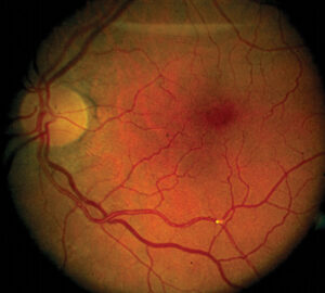
Patients report pain in the hands and feet, heat intolerance, skin lesions called angiokeratomas, and later organ failure (see retinal vascular tortuosity).
Corneal Findings in Fabry Disease
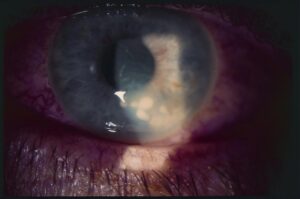
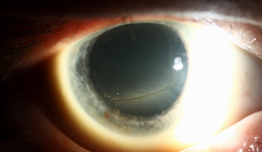
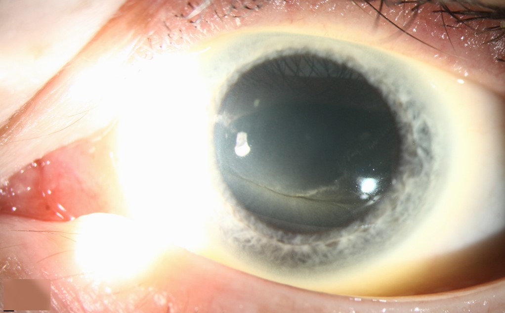
The classic sign is cornea verticillata, a whorl-like pattern of pale lines in the inferior epithelium.
It occurs in most males and many females, and it rarely affects vision.
Other drugs, such as amiodarone, cause a similar pattern, so the history matters.
Samiy and colleagues described its value as a clue to a treatable disease.
Fundus Explorer Pro
Photograph the retinal findings described here with the phone already in your pocket — 22 D optics and built-in illumination in one handheld unit.
From Choroida — the team behind this siteLens and Conjunctival Findings
Fabry cataract appears as a spoke-like opacity on the posterior lens capsule, with a granular anterior form.
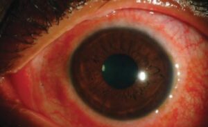
Conjunctival vessels may show tortuosity and aneurysmal dilatation.
These changes rarely affect vision, but they support the diagnosis.
Retinal Findings in Fabry Disease
- Tortuous retinal arterioles and venules
- Occasional vascular occlusions and optic neuropathy
- Retinal vessel tortuosity that correlates with disease severity
Sodi and colleagues reported ocular findings in a large survey and found vascular tortuosity in many patients.
Retinal changes may reflect the burden of vascular disease elsewhere.
When to Suspect and Test
Suspect Fabry disease in a young person with cornea verticillata and no drug exposure.
Also suspect it when a family history of kidney failure, stroke, or heart disease exists.
Measure alpha-galactosidase A activity in males, and perform genetic testing in all suspected patients, including females.
Management and Referral in Fabry Disease
Refer to a metabolic or nephrology clinic for treatment and family screening.
Ophthalmologists follow the eyes, treat cataract, and monitor for vascular events.
Treat other causes of the corneal pattern, and stop causative drugs when possible.
An Example Case
A 26-year-old man attends for blurred vision, and the slit lamp shows a whorl-like pattern in the inferior cornea.
He takes no amiodarone or other drugs known to cause the pattern.
He describes burning pain in his hands and episodes of heat intolerance.
Enzyme testing shows low alpha-galactosidase A activity, and genetics confirm a pathogenic variant.
His family undergoes screening, and he starts therapy under a metabolic team.
Questions Patients Ask
Patients ask whether the eye findings will cause blindness, and vision usually stays good.
They also ask whether the disease is hereditary, and the answer is yes, with X-linked inheritance.
Because relatives may be at risk, offer genetic counseling.
Pitfalls to Avoid
- Assuming that cornea verticillata always means a drug effect
- Overlooking the diagnosis in women, who may have mild or severe disease
- Ignoring retinal vessel tortuosity in a young patient with unexplained stroke
- Delaying referral when the family history suggests early kidney failure
Looking Ahead
New therapies, such as chaperones and gene therapy, are under study.
Also, newborn screening may identify patients earlier.
Therefore, ophthalmic findings will matter less for diagnosis but still support it.
Practical Points
Photograph the corneal pattern and the vessels for the record.
Then refer to a metabolic center for testing and management.
Also recommend screening of family members.
Cornea Verticillata Compared With Drug-Induced Patterns
Amiodarone, chloroquine, and other drugs produce a similar whorl.
Therefore, take a full drug history first.
Fabry disease usually presents earlier in life and with other systemic signs.
Also look for tortuous vessels and cataract as supporting signs.
Treatment in Brief for Fabry Disease
Enzyme replacement therapy and oral chaperone therapy slow disease progression in selected patients.
Early treatment gives the best results, so timely referral matters.
Also manage pain, kidney disease, and cardiovascular risk with the multidisciplinary team.
Family Screening in Fabry Disease
Because the disease is X-linked, mothers of affected sons are carriers, and daughters of affected men are carriers.
Therefore, offer testing to relatives.
Also advise genetic counseling before pregnancy.
Ophthalmic Follow-Up
Examine the cornea, lens, and retina every one to two years.
Then record any changes in tortuosity or cataract.
Also review the drug list, since new medicines may cause corneal changes.
Because Fabry disease affects many organs, share the findings with the care team.


Document what you see
Two smartphone imaging tools built for everyday clinic use — one for the slit lamp, one for the fundus.
From Choroida — the team behind this siteReferences
- Sodi A, Ioannidis AS, Mehta A, et al. Ocular manifestations of Fabry’s disease: data from the Fabry Outcome Survey. Br J Ophthalmol. 2007;91:210-214.
- Samiy N. Ocular features of Fabry disease: diagnosis of a treatable life-threatening disorder. Surv Ophthalmol. 2008;53:416-423.
- Zarate YA, Hopkin RJ. Fabry’s disease. Lancet. 2008;372:1427-1435.
- American Academy of Ophthalmology. Basic and Clinical Science Course, Section 12: Retina and Vitreous. AAO; current edition.

Writes and reviews the clinical articles on Educate. Every post is checked against peer-reviewed sources before it goes live.