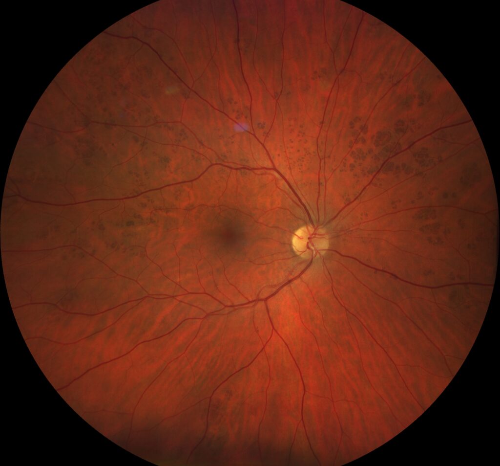
Polar bear tracks are clusters of flat, white or cream-colored spots at the level of the retinal pigment epithelium (RPE).
They are the unpigmented counterpart of grouped congenital hypertrophy of the RPE, and most are found by chance during a routine dilated examination. However, their pale color can mimic inflammatory or degenerative white lesions.
What Polar Bear Tracks Are
The finding is congenital and developmental, and clinicians also call it congenital grouped albinotic spots of the RPE. The arrangement resembles animal footprints, so the name stuck. Lesions usually occupy one sector of one eye, although bilateral cases have been reported, and because the cells are present from birth, the pattern generally stays stable for life.
Polar Bear Tracks on Fundus Examination
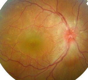
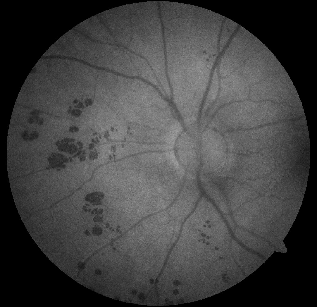
First, the examiner sees groups of small, round or oval white spots that are flat, sharply outlined and free of surrounding pigment. Spots often enlarge toward the periphery and then cluster like paw prints. The overlying retina and vessels look normal, and there is no vitreous cell, hemorrhage or retinal edema.
Fundus Explorer Pro
Photograph the retinal findings described here with the phone already in your pocket — 22 D optics and built-in illumination in one handheld unit.
From Choroida — the team behind this siteWhy the Spots Look White
Typical bear tracks look dark because their RPE cells carry dense melanin granules, whereas the cells in the white variant are thought to hold little or no melanin. As a result, the spots stand out as pale islands against the normal fundus, although the exact histologic basis has been studied far less than pigmented CHRPE.
Symptoms and Visual Function
Patients are almost always asymptomatic, and visual acuity is normal unless another disease coexists, while most clusters also lie outside the macula. Therefore, new blurring, photopsia or scotoma should prompt a search for another cause.
Fundus Autofluorescence
Autofluorescence is the most helpful single test, and the spots typically appear hypoautofluorescent, which fits reduced RPE fluorophore signal. Moreover, imaging can reveal more spots than ophthalmoscopy shows, and the pattern then stays unchanged on repeat images.
OCT Findings
OCT through a spot generally shows an intact inner retina, although subtle changes may appear in the outer retinal bands and the RPE line. However, there is no subretinal fluid, no elevation and no choroidal mass, and these negative findings help exclude active disease.
Polar Bear Tracks Versus Pigmented Bear Tracks
Both forms are grouped, sectoral and congenital, and both also follow the same benign course. The main visible difference is color, although mixed clusters containing pigmented and unpigmented spots have also been described. For the dark form and its imaging, see bear tracks, while solitary lesions behave differently, so review solitary CHRPE separately.
White Dot Syndromes: The Main Differential
MEWDS, APMPPE and birdshot chorioretinopathy also produce pale fundus spots. However, inflammatory white dots usually cause symptoms such as photopsia, blurring or blind spot enlargement, and they often show vitreous cells and change over weeks. Their angiographic and OCT signs are also distinctive (see white dot syndromes), whereas polar bear tracks are silent, fixed and sharply grouped.
Other White Lesions to Exclude
- Drusen, which lie beneath the RPE and favor the macula
- Fundus albipunctatus and retinitis punctata albescens, which cause night blindness
- Healed chorioretinal scars, which usually have pigmented borders
- Stargardt flecks, which are pisciform and centered on the posterior pole
- Paving stone degeneration, which shows bare sclera and large choroidal vessels
Polar Bear Tracks and Polyposis
Fundus lesions linked to familial adenomatous polyposis look different, and they are typically multiple, bilateral and oval, often with a depigmented halo or tail. Classic grouped CHRPE has not shown an association with polyposis, so a sectoral cluster alone does not call for a polyposis workup, although a positive family history deserves its own assessment.
Why Polar Bear Tracks Need No Treatment
The cells are benign and developmentally fixed, and no malignant potential has been established for grouped lesions. Laser, injections and supplements offer nothing here, and any intervention would therefore add risk without benefit.
Follow-Up for Polar Bear Tracks
First, record baseline wide-field color photographs and autofluorescence, then continue routine eye examinations at the usual interval. Repeat imaging if symptoms appear or the lesions seem to change. Finally, refer to ocular oncology for elevation, fluid, growth or a new nodule.
Counseling Patients With Polar Bear Tracks
Describe the finding as a birthmark in the layer beneath the retina, and explain that it is not inflammatory, infectious or cancerous. Family members do not need screening because of this finding alone, and give patients copies of their images for future examiners.
Pitfalls to Avoid
- Labeling the spots a white dot syndrome and starting steroids
- Ordering a full uveitis workup for fixed, silent lesions
- Confusing the pattern with polyposis-related fundus lesions
- Skipping baseline photographs and autofluorescence
- Missing a second disease when symptoms do appear


Document what you see
Two smartphone imaging tools built for everyday clinic use — one for the slit lamp, one for the fundus.
From Choroida — the team behind this siteReferences
- Kim DY, Hwang JC, Moore AT, Bird AC, Tsang SH. Fundus autofluorescence and optical coherence tomography of congenital grouped albinotic spots. Retina. 2010;30:1217-1222.
- Shields JA, Shields CL, Shah PG, et al. Lack of association among typical congenital hypertrophy of the retinal pigment epithelium, adenomatous polyposis, and Gardner syndrome. Ophthalmology. 1992;99:1709-1713.
- Traboulsi EI, Krush AJ, Gardner EJ, et al. Prevalence and importance of pigmented ocular fundus lesions in Gardner’s syndrome. N Engl J Med. 1987;316:661-667.
- Shields CL, Mashayekhi A, Ho T, Cater J, Shields JA. Solitary congenital hypertrophy of the retinal pigment epithelium: clinical features and frequency of enlargement in 330 patients. Ophthalmology. 2003;110:1968-1976.
- Fung AT, Pellegrini M, Shields CL. Congenital hypertrophy of the retinal pigment epithelium: enhanced-depth imaging optical coherence tomography in 18 cases. Ophthalmology. 2014;121:251-256.
Test yourself
A few questions straight from this article.
-
Polar bear tracks of the fundus are the unpigmented counterpart of which lesion?
Polar bear tracks, also called congenital grouped albinotic spots of the RPE, are the unpigmented counterpart of grouped CHRPE. -
What is the usual distribution of polar bear tracks in the fundus?
Lesions usually occupy one sector of one eye, although bilateral cases have been reported, and the pattern generally stays stable for life. -
Why do the spots of polar bear tracks look white rather than dark?
Pigmented bear tracks carry dense melanin granules, whereas cells in the white variant are thought to hold little or no melanin. -
How do polar bear track spots typically appear on fundus autofluorescence?
Autofluorescence is the most helpful single test; the spots appear hypoautofluorescent and imaging can reveal more spots than ophthalmoscopy. -
Which OCT finding fits polar bear tracks?
OCT shows an intact inner retina with at most subtle outer band changes, and no fluid, elevation or choroidal mass, which helps exclude active disease. -
Which feature favors a white dot syndrome over polar bear tracks?
MEWDS, APMPPE and birdshot usually cause symptoms, show vitreous cells and change over weeks, whereas polar bear tracks are silent and fixed. -
Which white fundus lesion differs from polar bear tracks by causing night blindness?
Fundus albipunctatus and retinitis punctata albescens cause night blindness, while polar bear tracks leave vision normal. -
Which fundus lesion pattern is linked to familial adenomatous polyposis, unlike polar bear tracks?
Polyposis-linked lesions are multiple, bilateral and oval, often with a halo or tail; a sectoral cluster alone does not call for polyposis workup. -
What management is appropriate for polar bear tracks?
The cells are benign and fixed, so treatment adds only risk; record baseline wide-field photographs and autofluorescence and continue routine examinations. -
Which change in a known polar bear track cluster warrants ocular oncology referral?
Repeat imaging if symptoms or change appear, and refer to ocular oncology for elevation, fluid, growth or a new nodule.

Writes and reviews the clinical articles on Educate. Every post is checked against peer-reviewed sources before it goes live.