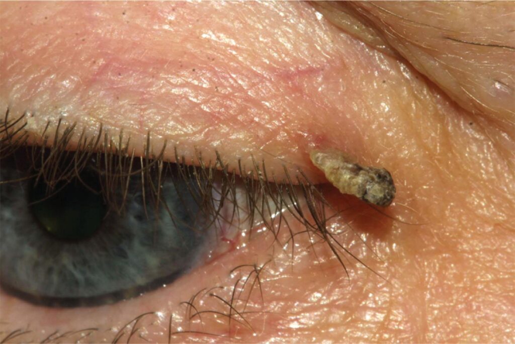
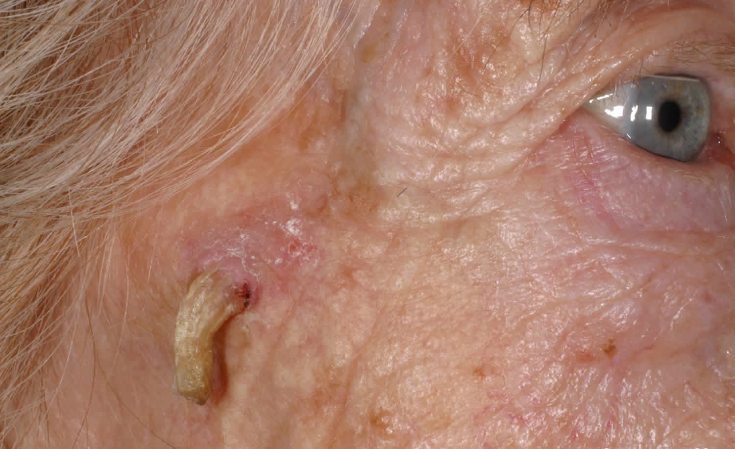
Cutaneous horn is a clinical description of a hard, conical projection of compacted keratin rising from the skin.
It is a sign, and the true diagnosis lies in the lesion at its base, which on the eyelid can be a harmless wart or an invasive carcinoma. Therefore, the horn tells you little until a pathologist examines the base.
What a Cutaneous Horn Is
The horn forms when an underlying lesion produces keratin faster than the surface can shed it, and the keratin then stacks into a firm, yellow-white or brown spike. Its length often exceeds its width and it may curve like a small animal horn, but the projection itself carries no diagnostic tissue.
Where Cutaneous Horn Appears on the Lid
Eyelid horns arise on the lid margin or on the skin of the anterior lamella, and patients are usually older adults with fair skin and long outdoor exposure. Horns can also arise on brow and canthal skin, where the same rules apply.
All-fit Slit-Lamp Adapter
Record and share exactly what you see at the slit lamp. One adapter fits any slit lamp or surgical microscope — and any smartphone.
From Choroida — the team behind this siteBenign Lesions at the Base
Many different benign lesions can produce a horn, for example:
- Seborrheic keratosis
- Verruca vulgaris (viral wart)
- Inverted follicular keratosis
- Trichilemmoma
- Epidermal inclusion cyst with keratin extrusion
Premalignant and Malignant Bases of a Cutaneous Horn
Actinic keratosis is a frequent premalignant base, and squamous cell carcinoma in situ and invasive squamous cell carcinoma also occur. Keratoacanthoma may also present with a keratin plug that forms a horn (see keratoacanthoma of the eyelid). Basal cell carcinoma is a rare base, although case reports describe it.
History in Cutaneous Horn
First, ask how long the horn has been present and whether it grew quickly. Then ask about bleeding, pain at the base, or regrowth after the patient picked it off, and a history of skin cancer, transplantation or immunosuppression also raises suspicion.
Examining the Base at the Slit Lamp
The slit lamp magnifies the junction between horn and skin very well, so look for induration, erythema, ulceration or pearly edges around the base. Next, check the margin for lash loss and distorted meibomian orifices, and finally feel the preauricular and submandibular nodes, because squamous cell carcinoma can spread there.
Features That Raise Concern
Clinical series suggest that some features favor a premalignant or malignant base.
- A broad base relative to horn height
- Induration or tenderness at the base
- Rapid growth over weeks
- Lash loss or margin notching
- Recurrence after previous removal
However, no single feature rules malignancy in or out.
Why Excision With Base Histology Matters
Excision with base histology is the only reliable way to name the lesion, because a shave of the horn alone removes keratin and leaves the diagnostic tissue behind. As a result, the pathologist may report only hyperkeratosis, and a carcinoma goes unrecognized. Therefore, include the base, a rim of normal skin, and enough depth to assess invasion.
Surgical Planning for a Cutaneous Horn
Small lesions away from the margin suit a simple elliptical excision, while margin lesions often need a full thickness pentagonal wedge to remove the base completely. When malignancy looks likely, plan margin control with frozen sections or Mohs surgery, and place an orienting suture so the pathologist can report each margin. Closure then follows the layered principles of eyelid laceration repair.
Reading the Pathology Report
A complete report names the base lesion, its margins and any invasion. Benign lesions with clear margins need no further surgery, and actinic keratosis calls for surveillance of the surrounding sun-damaged skin. However, invasive carcinoma with involved margins needs re-excision and possibly staging (see eyelid squamous cell carcinoma).
Cutaneous Horn Mimics
Some lid lesions look horn-like without a true keratin column, for example filiform warts and large papillomas that project from the margin. Dried crust over an ulcerated basal cell carcinoma can also resemble a horn, so soften and lift crust gently before you judge the base.
Counseling the Patient With a Cutaneous Horn
Many patients want the horn snipped off quickly for cosmetic reasons, so explain that the spike is only the surface, while the base holds the diagnosis. Most horns prove benign, so avoid alarming language, but state clearly that a minority hide skin cancer and that histology settles the question.
Follow-up After Excision of a Cutaneous Horn
Review the wound at one to two weeks for healing and margin alignment, then set the follow-up interval according to the histology. Patients with premalignant or malignant bases need periodic skin and lid examinations, and in addition you should advise hats, sunglasses and daily sun protection.
Pitfalls to Avoid
- Do not shave the horn flush and discard the base
- Never treat an unbiopsied horn with cryotherapy or cautery
- Do not ignore induration or lash loss at the base
- Avoid sending a suspicious specimen without orientation
- Do not forget regional lymph nodes in suspected carcinoma


Document what you see
Two smartphone imaging tools built for everyday clinic use — one for the slit lamp, one for the fundus.
From Choroida — the team behind this siteReferences
- Yu RC, Pryce DW, Macfarlane AW, Stewart TW. A histopathological study of 643 cutaneous horns. Br J Dermatol. 1991;124:449-452.
- Mencía-Gutiérrez E, Gutiérrez-Díaz E, Redondo-Marcos I, Ricoy JR, García-Torre JP. Cutaneous horns of the eyelid: a clinicopathological study of 48 cases. J Cutan Pathol. 2004;31:539-543.
- Copcu E, Sivrioglu N, Culhaci N. Cutaneous horns: are these lesions as innocent as they seem to be? World J Surg Oncol. 2004;2:18.
- American Academy of Ophthalmology. Basic and Clinical Science Course, Section 7: Oculofacial Plastic and Orbital Surgery. San Francisco: AAO; 2023.
Test yourself
A few questions straight from this article.
-
What is an eyelid cutaneous horn, diagnostically speaking?
A cutaneous horn is a clinical description of compacted keratin; the true diagnosis lies in the lesion at its base, from wart to invasive carcinoma. -
How does a cutaneous horn form on the eyelid?
An underlying lesion produces keratin faster than the surface can shed it, so the keratin stacks into a firm spike that itself carries no diagnostic tissue. -
Which premalignant lesion is a frequent base of an eyelid cutaneous horn?
Actinic keratosis is a frequent premalignant base, and squamous cell carcinoma in situ and invasive squamous cell carcinoma also occur. -
Which feature of an eyelid cutaneous horn favours a premalignant or malignant base?
A broad base, induration or tenderness, rapid growth, lash loss and recurrence favour a worrying base, though no single feature rules malignancy in or out. -
Why does shaving an eyelid cutaneous horn flush with the skin risk a missed carcinoma?
A shave removes only keratin, so the pathologist may report hyperkeratosis alone; excision should include the base, a rim of normal skin and enough depth to assess invasion. -
Which excision usually suits a cutaneous horn arising on the eyelid margin?
Small lesions away from the margin suit an elliptical excision, but margin lesions often need a full-thickness pentagonal wedge to remove the base completely. -
Which lymph nodes should be palpated when examining an eyelid cutaneous horn?
After examining the base and margin, the preauricular and submandibular nodes should be felt, because squamous cell carcinoma can spread there. -
What follow-up does actinic keratosis at the base of an excised eyelid horn call for?
Benign lesions with clear margins need no further surgery, actinic keratosis calls for surveillance of the sun-damaged skin, and invasive carcinoma with involved margins needs re-excision. -
Which lesion can mimic an eyelid cutaneous horn with dried crust rather than a keratin column?
Dried crust over an ulcerated basal cell carcinoma can resemble a horn, so crust should be softened and lifted gently before judging the base. -
Which patients typically develop eyelid cutaneous horns?
Eyelid horns arise on the margin or anterior lamella skin, usually in older adults with fair skin and long outdoor exposure.

Writes and reviews the clinical articles on Educate. Every post is checked against peer-reviewed sources before it goes live.